


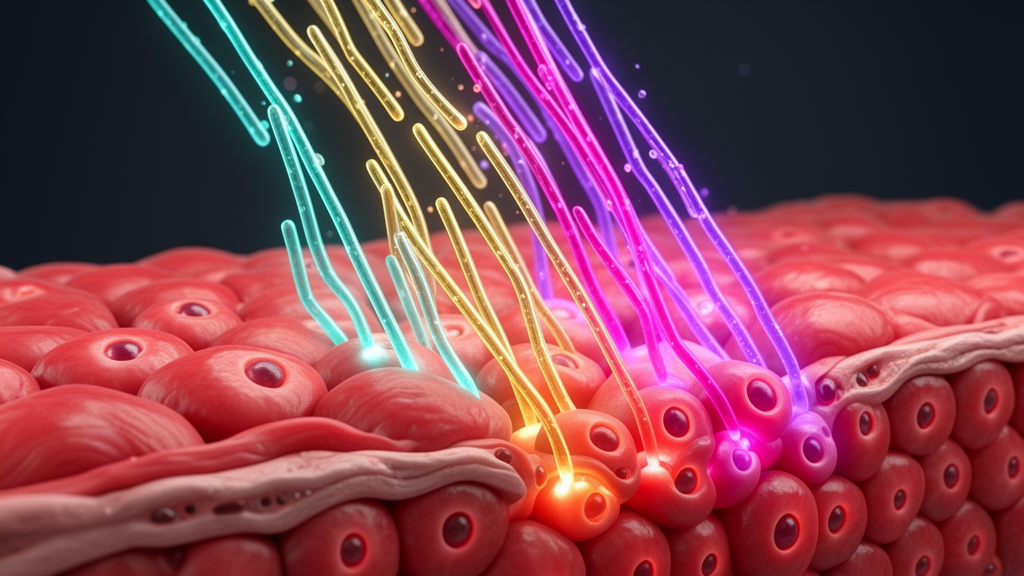
When a mammalian heart suffers a myocardial infarction, ischemia and hypoxia kill large numbers of cardiomyocytes, the contractile cells that make up the beating muscle. The dead tissue is replaced by a fibrotic scar, an irreversible loss that compromises pumping function and sets the stage for progressive heart failure. Current therapies such as revascularization, beta-blockers and ACE inhibitors can slow this decline, but none of them regenerate the lost muscle cells or reverse fibrosis. A comprehensive review published in Cell Regeneration examines the molecular machinery that governs whether a heart repairs itself or simply scars, and asks how that machinery might be harnessed therapeutically.
The answer, the review argues, lies in a set of evolutionarily conserved signaling pathways: Hippo-YAP, Wnt/beta-catenin, neuregulin 1 (NRG1)-ErbB, mitogen-activated protein kinase (MAPK) and Notch. These networks orchestrate cardiomyocyte proliferation, cell death, metabolic adaptation and remodeling of the extracellular matrix, and their activity differs dramatically between species. Fish, amphibians and even neonatal mammals such as mice and pigs can regenerate heart muscle robustly after injury, through cardiomyocyte dedifferentiation, proliferation and redifferentiation. Adult mammalian cardiomyocytes, by contrast, exit the cell cycle and suppress pro-regenerative signaling, which is why human hearts cannot rebuild themselves after a heart attack.
The Hippo pathway emerges as one of the most compelling targets. This organ-size control system, built around the kinase cascade MST1/2 and LATS1/2 and the downstream effectors YAP and TAZ, normally holds cardiomyocyte proliferation in check. When the pathway is inactivated, unphosphorylated YAP accumulates in the nucleus and switches on genes for cell division and survival. In mice, cardiomyocyte-specific deletion of Sav1 or Lats1/2 drives cell cycle re-entry and improves functional recovery after infarction or apical resection. YAP activation upregulates genes involved in cytoskeletal remodeling, suggesting that structural reorganization, not just proliferation, contributes to repair. Conversely, fetal inactivation of Yap1 causes lethal myocardial hypoplasia, underscoring how central this effector is to heart development.
Therapeutic strategies built on YAP are advancing quickly. Adeno-associated virus serotype 9 delivery of a phosphorylation-resistant YAP S127A mutant reduces scarring and promotes new blood vessel growth after infarction in mice, while sustained expression of human YAP reactivates proliferation in adult cardiomyocytes. A constitutively active mutant called YAP5SA induces adult cardiomyocyte division but also triggers Hippo-mediated negative feedback that prevents excessive cell division, an apparent built-in safety valve. More recently, a modified mRNA system and a drug-elicitable alternative-splicing module have enabled transient, controllable YAP5SA delivery, designed to stimulate regeneration while limiting off-target risks. Post-translational regulation adds another layer: after infarction, acetylation of YAP at lysine 265 sequesters the protein in the cytoplasm on microtubules, and a mutation preventing this modification restores nuclear localization and regenerative capacity.
Wnt/beta-catenin signaling presents a more complicated picture, one the review describes as remarkably context-dependent. In neonatal mice and human stem cell-derived cardiomyocytes, canonical Wnt signaling promotes proliferation, but in the adult mouse heart it instead provides cardioprotection while preventing reactivation of the pro-proliferative gene program. Consistent with this, small-molecule inhibitors of canonical Wnt signaling promote regeneration in adult hearts, and in zebrafish, Wnt inhibitors secreted from the epicardium, fibroblasts and myocardium mediate regeneration after apex resection. Yet other studies show that activating Wnt signaling, for example by genetically ablating the beta-catenin-destroying kinase GSK3beta or treating cells with the inhibitor CHIR99021, also enhances cardiomyocyte proliferation and post-infarction remodeling. The review attributes these apparent contradictions to developmental stage, pathway antagonism between canonical and non-canonical branches, crosstalk with other pathways, and effects on non-muscle cells such as endothelial cells and macrophages.
The NRG1-ErbB axis offers a cleaner story. NRG1, an epidermal growth factor family member, binds ErbB4, which heterodimerizes with ErbB2 and triggers PI3K/AKT and MAPK/ERK signaling. In adult zebrafish, Nrg1 robustly activates cardiomyocyte mitosis, and in neonatal mammals the ErbB2 receptor is transiently re-expressed after injury, downregulating mature cardiomyocyte markers and upregulating embryonic genes. This transient regenerative program fades early in postnatal life, but it can be rescued by recombinant NRG1 or constitutively active ErbB2, which promotes cell cycle re-entry in the injured zone. Single-cell transcriptomics has revealed that NRG1/ErbB2 drives a glycolytic metabolic shift in proliferating border-zone cardiomyocytes, a finding later confirmed in the mouse heart. NRG1 also counteracts oxidative stress-induced apoptosis and, in remodeling and heart failure models, recombinant NRG1beta attenuates cardiac fibrosis.
MAPK signaling plays dual roles that depend on which family member is considered. p38 MAPK acts as a brake: its active form suppresses mitosis-associated genes, and pharmacological inhibition or knockdown of p38alpha improves ventricular function and reduces fibrosis. ERK, in contrast, is a central pro-regenerative hub. In zebrafish, injury-generated hydrogen peroxide inactivates the phosphatase Dusp6, relieving inhibition of ERK1/2 and synergizing with immune cell recruitment to drive repair. ERK also amplifies ErbB2 signaling through positive feedback, and diverse upstream inputs, including the extracellular matrix protein Agrin binding to dystroglycan, converge on ERK and YAP to promote dedifferentiation and proliferation. Notch signaling, meanwhile, is activated in the endocardium and epicardium after zebrafish ventricular injury and integrates mechanical cues: hemodynamic changes sensed by mechanosensitive channels and primary cilia induce klf2a, which activates Notch and non-cell-autonomously initiates myocardial ErbB2 and BMP signaling. In adult mammals, however, permanent epigenetic modifications silence the Notch response, constraining the pathway’s regenerative potential.
Beyond these core pathways, the review highlights the cellular microenvironment as an active participant in repair. The extracellular matrix provides mechanical support and dynamically regulates inflammatory responses, proliferation and migration. Biomimetic strategies exploit this: decellularized neonatal mouse matrix softens tissue stiffness to activate YAP signaling and enhance angiogenesis, injectable cardiac-derived matrix particles improve function in murine infarction models, and porcine small intestinal submucosal matrix combined with human cardiovascular progenitor cells synergistically enhances graft survival. Individual matrix proteins carry distinct regenerative signatures. Agrin displaces YAP from the dystrophin glycoprotein complex, reinitiating cell cycle progression. FSTL1, periostin and versican modulate fibroblast activity, collagen deposition and cardiomyocyte proliferation with context-dependent effects, and growth factors including PDGF, FGF, VEGF, IGF and the myeloid-derived growth factor each shape repair through specific receptor pathways, sometimes with fibrotic side effects that demand careful isoform-specific targeting.
Metabolism emerges as a master switch. After birth, cardiomyocyte energy metabolism shifts from glycolysis to fatty acid beta-oxidation, accompanied by rising mitochondrial reactive oxygen species that damage DNA and enforce cell cycle arrest. Experimental disruption of fatty acid oxidation, sustained systemic hypoxia at 7 percent oxygen, or overexpression of the glucose transporter GLUT1 can partially reverse this maturation and reactivate proliferation. Transcription factors coordinate these programs: Meis1 and Hoxb13 enforce cell cycle arrest in postnatal cardiomyocytes, and their dual deletion in adult mice enhances proliferation and improves post-infarction contractility, with small-molecule inhibitors of the Meis1-DNA interaction showing benefit in preclinical models of mice and pigs. GATA4, TBX20, E2F family members and PITX2 add further layers, linking proliferation, mitochondrial health and redox balance. Cell cycle proteins themselves are therapeutic levers: co-overexpression of cyclins and CDKs increases proliferation in mouse and human cardiomyocytes, and transient cyclin D2 delivery via modified mRNA improves ventricular function in large animal infarction models.
The review closes by surveying the technologies now accelerating the field. Single-cell and spatial transcriptomics have generated regeneration atlases and identified conserved regulators such as angpt4, while high-content screening and molecular dynamics simulations have surfaced candidate drugs including nimodipine and salvianolic acid B. Engineered heart muscle grafts from induced pluripotent stem cell-derived cardiomyocytes have shown six-month retention and functional recovery in non-human primates, left ventricular assist devices have been reported to enhance cardiomyocyte renewal rates sixfold, and Hippo-targeted gene therapy is approaching clinical testing supported by large animal models. Yet the authors are candid about the barriers: undefined spatiotemporal thresholds for clinically meaningful regeneration in adult human hearts, delivery challenges including off-target effects and immunogenicity, pathophysiological gaps between murine models and human disease, and unresolved links between metabolic reprogramming, epigenetics and regenerative outcomes. Bridging those gaps, they argue, will require integrating multi-omics insights, smart biomaterials and interdisciplinary approaches to move from transient repair toward true functional reconstruction of the injured heart.
Subject of Research: Signaling pathways regulating cardiac regeneration after myocardial injury
Article Title: Signaling pathways regulating cardiac regeneration
Article References: Li, J., Hu, M., Ding, L., & Wang, Y. (2026). Signaling pathways regulating cardiac regeneration. Cell Regeneration, 15(1), Article 24. https://doi.org/10.1186/s13619-026-00297-7
Image Credits: AI Generated
DOI: 10.1186/s13619-026-00297-7
Keywords: cardiac regeneration, cardiomyocyte proliferation, Hippo-YAP, Wnt signaling, NRG1-ErbB, Notch, myocardial infarction, metabolism, transcription factors, extracellular matrix, heart failure, gene therapy
News Source: Gregory Coleman. (October 28, 2026). How Hippo, Wnt, NRG1 and Notch pathways could unlock heart regeneration. Scienmag.



