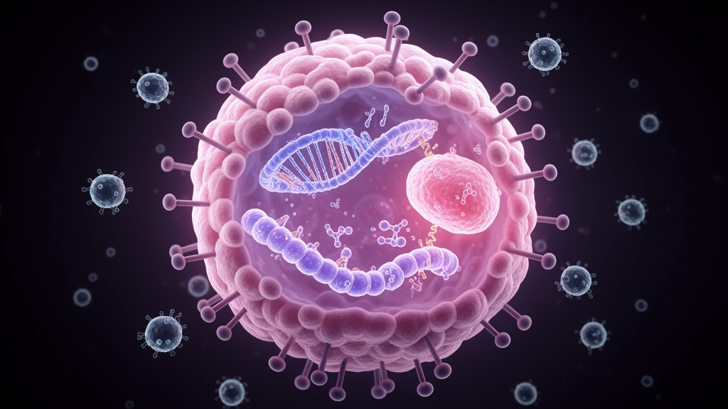
Colorectal cancer remains a leading cause of cancer-related mortality worldwide, and a new study published in Cell Biology and Toxicology points to a single molecule, cadherin 3 (CDH3), as a hub connecting tumor aggressiveness with the cancer’s ability to hide from the immune system. The work, led by Chunhui Jiang, Danping Shen, Ye Liu and Lei Gu, reports that CDH3 is kept at high levels in colorectal cancer cells through two independent mechanisms: a chemical modification of its messenger RNA installed by the enzyme NAT10, and the removal of ubiquitin tags from its protein by the deubiquitinase USP19.
The team began with an integrative bioinformatics search for key biomarkers, combining weighted gene co-expression network analysis with machine learning algorithms. That screen pinpointed CDH3, which the researchers found significantly upregulated in colorectal cancer tissues and cells compared with controls, a difference that reached statistical significance at P < 0.05. Importantly, higher CDH3 was also correlated with adverse patient prognosis, marking it as a molecule associated with worse clinical outcomes.
To probe what CDH3 actually does, the researchers silenced the gene in colorectal cancer cells in the laboratory. Loss of CDH3 suppressed three hallmark malignant behaviors: cell proliferation, migration, and angiogenesis, the process by which tumors coax new blood vessels into forming. Each of these functional readouts changed significantly, with P < 0.05 reported across the assays of cell viability, proliferation, migration and tube formation.
Using co-culture systems in which colorectal cancer cells were grown alongside peripheral blood mononuclear cells, CD8+ T cells, and THP-1-derived macrophages, the team measured how CDH3 shapes the tumor’s immune microenvironment. When CDH3 was knocked down, CD8+ T cells underwent less apoptosis, PBMCs secreted higher amounts of the inflammatory cytokines tumor necrosis factor-alpha and interferon-gamma, and M2 macrophage polarization was inhibited. All of these effects were statistically significant.
Mechanistically, the study disentangled two layers of CDH3 regulation. At the RNA level, the acetyltransferase NAT10 deposited N4-acetylcytidine (ac4C) modifications on CDH3 messenger RNA, which stabilized the transcript, as confirmed by ac4C RNA immunoprecipitation alongside RNA pull-down and dual-luciferase reporter assays. At the protein level, ubiquitin specific peptidase 19 (USP19) physically interacted with CDH3 and removed K48-linked ubiquitin chains, the molecular marks that normally condemn a protein to destruction by the proteasome. Co-immunoprecipitation and ubiquitination assays supported this interaction, meaning USP19 prevents CDH3 protein degradation.
The researchers then tested whether restoring CDH3 could rescue the damage done by removing either upstream regulator. In both cell culture and animal models, overexpressing CDH3 partially reversed the suppressive effects of NAT10 or USP19 knockdown on malignant phenotypes and on in vitro immune escape. In vivo experiments using HCT-116 cells in tumorigenicity and lung metastasis models showed the same pattern: CDH3 overexpression partially rescued the reduced tumor growth and metastatic colonization caused by knocking down NAT10 or USP19. This rescue experiment is the strongest evidence in the study that CDH3 sits downstream of both regulators and functions as their common effector.
The dual-regulation architecture is notable because it means CDH3 abundance is reinforced at two separate points: NAT10 keeps the mRNA template plentiful and long-lived, while USP19 keeps the resulting protein from being recycled. Disrupting either arm lowered CDH3 enough to weaken tumor growth, spread, and immune evasion in the reported models, suggesting that the NAT10/USP19-CDH3 axis could be targeted at multiple nodes.
The authors are careful about the limits of their evidence. The immune experiments relied on co-cultures with human immune cell lines and PBMCs rather than an intact immune system, and the in vivo work used standard xenograft-style models with HCT-116 cells. The conclusion explicitly states that targeting the NAT10/USP19-CDH3 axis represents a promising candidate therapeutic strategy for colorectal cancer, although further validation in immunocompetent models is warranted. In other words, whether these mechanisms hold in animals with functioning immune systems, and whether they can be drugged safely in patients, remains to be demonstrated.
Even with those caveats, the study adds CDH3 to a short list of molecules whose cancer-promoting roles span both intrinsic malignancy and immune suppression. If follow-up work in immunocompetent models confirms the findings, the ac4C-deubiquitination partnership described here could offer diagnostic markers for prognosis and, potentially, combination targets designed to strip colorectal tumors of both their growth advantage and their immunological camouflage.
Subject of Research: Dual post-transcriptional and post-translational regulation of CDH3 in colorectal cancer progression and immune evasion
Article Title: Dual regulation of CDH3 by NAT10-mediated ac4C modification and USP19-mediated deubiquitination promotes colorectal cancer progression and immune evasion
Article References: Jiang, C., Shen, D., Liu, Y., & Gu, L. (2026). Dual regulation of CDH3 by NAT10-mediated ac4C modification and USP19-mediated deubiquitination promotes colorectal cancer progression and immune evasion. Cell Biology and Toxicology. https://doi.org/10.1007/s10565-026-10325-6
Image Credits: AI Generated
DOI: 10.1007/s10565-026-10325-6
Keywords: colorectal cancer, CDH3, NAT10, ac4C modification, USP19, deubiquitination, immune evasion, CD8+ T cells, M2 macrophage polarization, biomarker, mRNA stability, Dual
News Source: Nathaniel Bowman. (October 29, 2026). NAT10 and USP19 jointly stabilize CDH3 to drive colorectal cancer growth and immune evasion. Scienmag.