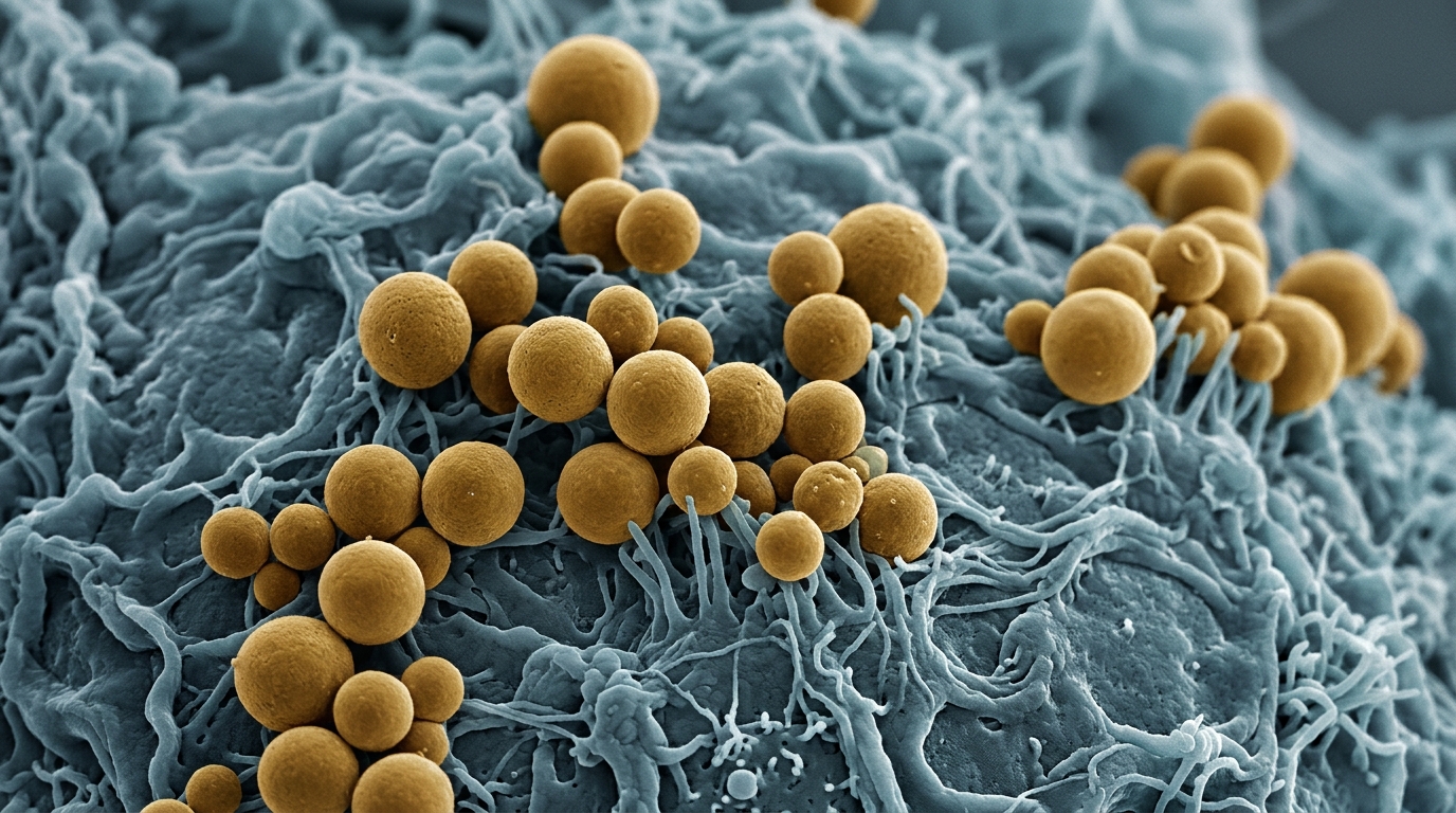
Nanoparticle-based carriers for chemotherapy have shown markedly better antitumor effects than free drugs in animal models of head and neck cancer, according to a systematic review published in Cancer Nanotechnology. The review, led by Omnia M. Abdallah, Marwa Mokbel Elshafei, and Hadeel Ahmad Kotat, pulled together 14 preclinical studies in mice, rats, and guinea pigs that compared chemotherapeutic drugs loaded onto nanomaterials with the same drugs given alone.
The authors searched PubMed, Web of Science, and Science Direct through 14 May 2024, following PRISMA 2020 guidelines and registering the protocol in PROSPERO under CRD42024512872. After removing duplicates, the keyword strategy yielded 873 studies, which the team screened down to the 14 included articles. Each study had to use an animal model, load a chemotherapeutic drug onto a nanomaterial, and include a control group receiving either the free drug or the nanomaterial alone. In vitro work, crossover designs, case reports, and studies without controls were excluded.
The included studies tested a broad range of nanocarrier forms, including nanosheets, nanomicelles, nanoshells, and nanospherical carriers, delivering chemotherapeutic drugs such as cisplatin, doxorubicin, dasatinib-oxaliplatin, and gefitinib. Another used hollow mesoporous silica nanoparticles carrying doxorubicin and paclitaxel, where the combination formulation achieved complete tumor growth suppression in mice treated weekly for three weeks.
Targeted delivery systems also showed strong results, with polymeric micelles carrying cisplatin and modified with a cyclic Arg-Gly-Asp peptide accumulating rapidly in tumors. Polymeric micelles carrying cisplatin, modified with a cyclic Arg-Gly-Asp peptide on their surface, accumulated rapidly in tumors, reaching more than 3 percent of the injected dose per gram within one hour and maintaining high levels for 24 hours. These targeted micelles suppressed lymphatic metastasis more effectively than free cisplatin, with the strongest tumor suppression and extended survival in the animal models. In a separate study, polymeric micelles carrying cisplatin reduced the incidence of lymphatic metastasis to 1 in 8 mice, compared with 7 in 8 in untreated controls and 3 in 8 in mice given free cisplatin.
Several formulations exploited the acidic, oxygen-poor tumor microenvironment, including hollow manganese dioxide nanoshells that decomposed hydrogen peroxide in the tumor microenvironment to release oxygen and relieve hypoxia. Hollow manganese dioxide nanoshells carrying cisplatin and docetaxel decomposed hydrogen peroxide in the tumor microenvironment, releasing oxygen and relieving hypoxia, which is known to make cancer cells more aggressive and resistant to chemotherapy. These nanoshells slowed tumor growth by day 28 and showed efficient renal clearance with no apparent toxicity. Another study used macrophage membrane-coated doxorubicin nanoparticles, hypothesizing that the membrane would burst at the acidic pH inside tumors, releasing the drug only within cancer cells. These coated particles evaded immune clearance, homed to tumors, and penetrated deeper than uncoated counterparts.
Toxicity findings favored the nanoparticle approaches across the board. Nanoparticle-treated groups maintained stable body weight and showed minimal organ toxicity, whereas free drug groups exhibited weight loss and systemic toxicity. Free doxorubicin caused myocardial fiber disruption and nuclear deformation in mouse hearts, while the nanoparticle formulations preserved normal cardiac architecture. Polymeric micelles carrying cisplatin reduced serum creatinine levels compared with free cisplatin, indicating less kidney damage. Free gefitinib was rapidly cleared from plasma within two hours, whereas lipid-polymer hybrid nanoparticle formulations sustained plasma retention until 48 hours, with higher drug concentrations in tumor tissue and lower concentrations in the heart and kidney.
Histopathological examinations reinforced these patterns. Treated tumors showed increased necrosis, apoptosis, and reduced proliferation, measured by decreased Ki67 expression. Tumor sections from mice given the manganese dioxide nanoshells showed extensive tumor cell damage, while liver and kidney tissues showed no histological evidence of toxicity four weeks after treatment. In one study, bilirubin-coated gold nanoparticles enhanced the expression of pro-apoptotic proteins including p53, Bax, caspase-3, and caspase-9, and downregulated the anti-apoptotic protein Bcl-2 in tumor tissue more effectively than bilirubin alone, suggesting a shift toward apoptosis induction in multidrug-resistant oral carcinoma xenografts.
The review also flagged important methodological weaknesses. Risk of bias was assessed using the SYRCLE tool, generating 140 entries across the 14 studies. Of these, 39.28 percent were answered yes, 52.85 percent were unclear, and 7.85 percent were no. Only six studies showed a moderate risk of bias with no high-risk items. The authors noted that the majority of studies had unclear or limited details on randomization methods and blinding, and that tumor size was typically measured with calipers, an operator-dependent method susceptible to measurement bias when assessors are not blinded to treatment allocation. Consequently, the reported tumor inhibition effect sizes may be overestimated and should be interpreted with caution.
The authors also cautioned that differences in dosing strategies across studies may have influenced treatment comparisons. Enhanced bioavailability and targeted delivery could result in higher exposure efficacy at equivalent doses, meaning the apparent superiority of nanomaterials should be cautiously interpreted. They called for equitoxic dose comparisons in future studies to more accurately assess the therapeutic advantage of nanomaterials carrying chemotherapeutic drugs. Additionally, the dominance of subcutaneous xenograft models, which represent localized tumor growth and lack key features of the native tumor microenvironment, may overestimate treatment efficacy and not fully predict clinical performance.
Overall, the review concluded that nanomaterial-based chemotherapeutic delivery systems positively affected tumor growth rates, toxicity, metastasis, and histopathological findings in the included preclinical studies. The authors emphasized that these findings warrant urgent further investigation, particularly regarding the safety and efficacy of this form of therapy, before any acceleration toward clinical trials. They noted that the number of included studies was low, indicating the need for further in-depth studies covering mechanistic insights and advanced animal models to better understand which nanomaterials work best with specific chemotherapeutic agents against particular tumor types.
Subject of Research: Preclinical efficacy of nanomaterial-based chemotherapeutic delivery in head and neck cancer animal models
Article Title: Efficacy of nanomaterials in the treatment of head and neck cancer: a systematic review
Article References: Abdallah, O. M., Elshafei, M. M., & Kotat, H. A. (2026). Efficacy of nanomaterials in the treatment of head and neck cancer: a systematic review. Cancer Nanotechnology, 17(1), Article 46. https://doi.org/10.1186/s12645-026-00413-x
Image Credits: AI Generated
DOI: 10.1186/s12645-026-00413-x
Keywords: nanoparticles, head and neck cancer, chemotherapy delivery, systematic review, preclinical animal models, tumor growth inhibition, reduced toxicity, metastasis suppression, cisplatin, doxorubicin, polymeric micelles, mesoporous silica
News Source: Nathaniel Bowman. (November 5, 2026). Nanoparticle drug carriers curb tumor growth and toxicity in head and neck cancer animal studies. Scienmag.