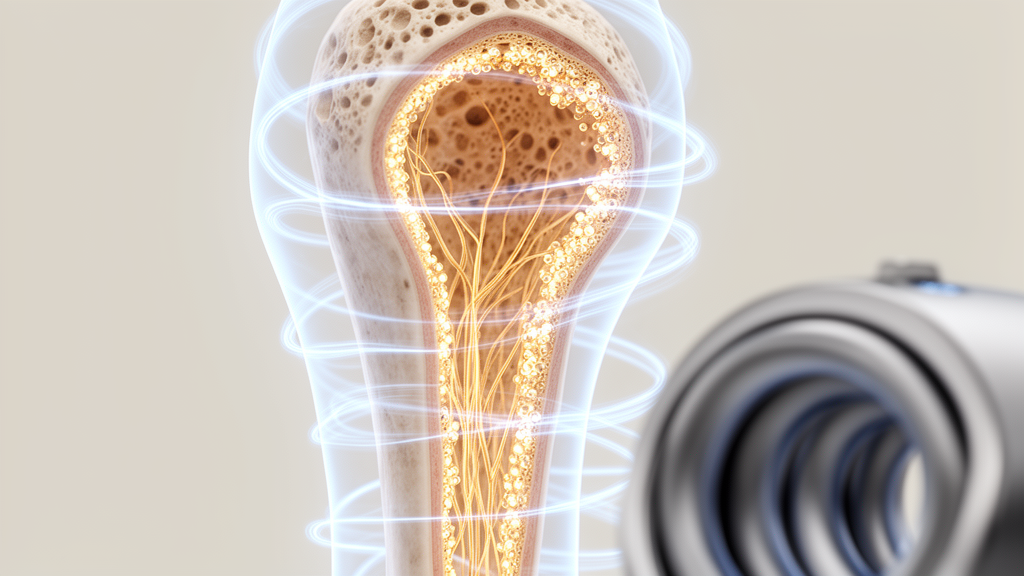
Chronic stress has long been known to hollow out the skeleton, but the precise neural machinery that translates psychological strain into fragile bones has remained frustratingly opaque. Now a study published in Nature Communications maps, in remarkable detail, an unexpected answer: a self-contained neural circuit buried inside the bone marrow itself, which can be switched by pulsed electromagnetic fields to protect the skeleton from the ravages of sustained stress. The work, led by Tiantian Wang and Zhen Hong of West China Hospital, Sichuan University, identifies sympathetic neuropeptide Y as the dominant pathological driver of stress-induced osteoporosis and demonstrates that a non-invasive physical therapy works by silencing this molecule through a local sensory-to-sympathetic feedback loop that operates entirely without the brain.
The clinical stakes are considerable. Meta-analyses have firmly established depression as a major risk factor for osteoporosis: people with depression show reductions of 7 to 15 percent in bone mineral density at clinically critical sites such as the spine and hip, and more than 40 percent of these patients develop the disease. Unlike postmenopausal osteoporosis, stress-induced bone loss strikes premenopausal women and adolescents, potentially condemning them to lifelong skeletal fragility, a burden compounded by the recurrent nature of depressive episodes. While recent studies have traced central brain circuits that mediate stress-induced bone loss, the specific efferent molecules and the possibility of targeting them non-invasively remained unresolved questions at the heart of the new investigation.
The researchers began with an unpredictable chronic mild stress model in male mice, an eight-week regimen of randomized daily stressors including restraint, cage dampening, and elevated-platform exposure. As expected, stressed mice developed anxiety-like behaviors and significant trabecular bone loss in the femur, with reduced bone volume fraction, fewer trabeculae, and wider separation between them. Cortical bone deteriorated in parallel. Daily treatment with pulsed electromagnetic fields, delivered for one hour per day over four weeks at 8 hertz and 3.8 millitesla, largely prevented this deterioration in both the trabecular and cortical compartments. Crucially, the therapy restored bone formation rates, measured by calcein double-labeling, and reversed a pathological shift in the marrow toward fat accumulation at the expense of bone-building osteoblast activity.
The cellular target of this protection proved to be a surprising one: bone marrow adipocytes, the fat cells that populate the skeletal interior. In stressed mice, these adipocytes showed hallmark signs of cellular senescence, including elevated DNA damage marked by γH2AX and increased senescence-associated β-galactosidase activity. Pulsed electromagnetic field treatment effectively cleared this senescent burden. Yet when the team exposed isolated adipocytes directly to the electromagnetic fields in a dish, nothing happened, indicating that the effect is indirect and requires an intermediary cell type. That intermediary, the experiments revealed, is the sympathetic nervous system, whose nerve fibers course through the marrow in close anatomical proximity to sensory nerves, a spatial arrangement confirmed by three-dimensional imaging of optically cleared bone.
Using high-resolution proteomic analysis of the sympathetic neuron secretome, the researchers found that neuropeptide Y, a canonical sympathetic neurotransmitter, was among the most significantly downregulated secreted proteins after pulsed electromagnetic field treatment. The finding was validated at both protein and transcript levels. Causality was established through a series of decisive experiments: recombinant neuropeptide Y alone was sufficient to drive senescence markers such as Cdkn1a and Cdkn2a in adipocytes, adding it back to conditioned medium from treated neurons restored the pro-senescent activity, and, most strikingly, transplanting adipocytes that had been pre-exposed to neuropeptide Y into healthy young recipient mice induced genuine bone loss, marrow fat expansion, and suppressed bone formation. Neuropeptide Y, in other words, does not merely correlate with the pathology; it actively manufactures it.
Genetic proof followed. Mice engineered to lack neuropeptide Y specifically in tyrosine hydroxylase-expressing sympathetic neurons were completely protected from chronic stress-induced bone loss, exhibiting preserved bone mass, higher mineral apposition rates, reduced marrow adiposity, and fewer senescent adipocytes. In a further twist, these knockout mice were also fully resistant to bone loss induced by chronic isoproterenol infusion, a pharmacological clamp that maximally stimulates β-adrenergic signaling. This positions neuropeptide Y, rather than the classical neurotransmitter norepinephrine, as the dominant pathological effector of sympathetic bone catabolism, a genuine paradigm shift in how scientists understand the sympathetic control of the skeleton under sustained pathological stress.
The upstream arm of the circuit proved equally elegant. Pulsed electromagnetic fields act on sensory nerves within bone, which respond by locally releasing semaphorin 3A, a secreted protein best known for guiding axons during development but increasingly recognized as a potent osteoanabolic factor. In wild-type mice, chronic stress depleted bone marrow semaphorin 3A, and the therapy robustly restored it, while serum levels remained untouched, indicating a strictly local mode of action. When sensory neurons were genetically ablated, every therapeutic effect of the electromagnetic fields vanished: bone mass was not rescued, sympathetic tone was not normalized, adipocyte senescence persisted, and local semaphorin 3A remained low. Conversely, injecting recombinant semaphorin 3A directly into the femur of sensory nerve-ablated mice rescued trabecular bone, proving the molecule both necessary and sufficient downstream of sensory nerves.
Perhaps the most audacious experiment was a complete spinal cord transection at the tenth thoracic level, physically severing all connections between the brain and the hindlimbs. Even in these animals, pulsed electromagnetic field treatment remained fully effective, suppressing sympathetic tone, activating sensory nerves, and reducing senescent adipocyte burden. Combined with the observation that systemic stress markers such as corticosterone and inflammatory cytokines were not normalized by the therapy, and that anxiety-like behaviors persisted, the evidence converges on a striking conclusion: this is a form of peripheral interoception, a tissue-autonomous neural reflex in which sensory nerves directly perceive the physical stimulus and fine-tune neighboring sympathetic terminals through semaphorin 3A, all without any central nervous system integration. The circuit challenges the conventional assumption that sensory-sympathetic communication must be routed through the brain or spinal cord.
The receiving end of the pathological signal also came into focus. Neuropeptide Y acts predominantly through the Y1 receptor, and double immunofluorescence localized this receptor to bone marrow adipocytes after stress. Mice lacking Y1R specifically in adipocytes were completely resistant to stress-induced bone loss, and pulsed electromagnetic fields conferred no additional benefit upon them, while a pharmacological Y1R agonist completely abrogated the therapy’s osteoprotective effect in wild-type mice. In vitro, recombinant neuropeptide Y triggered DNA damage and senescence programs in adipocytes through Y1R, effects blocked by the selective antagonist BIBO3304. Translational support came from re-analysis of a public human transcriptomic dataset, which revealed significantly elevated Y1R expression in osteoporotic bone, with concordant trends toward increased NPY and decreased SEMA3A, mirroring the murine signature and suggesting the mechanism is conserved in human disease.
The study also reframes what pulsed electromagnetic fields actually are. Rather than a generic osteogenic stimulus, the therapy emerges as a physiological rheostat that recalibrates local neural balance within bone. In aged mice, where sympathetic activity is pathologically diminished, the fields restore it; in chronically stressed mice, where sympathetic tone is pathologically elevated, they suppress it. In both contexts, sympathetic nerve density is steered back toward a physiological setpoint, consistent with a homeostatic modulator that harnesses innate circuitry for state-dependent therapy. The biophysical trigger likely involves direct depolarization of sensory neurons through L-type voltage-gated calcium channels, which couples neuronal activity to SNARE-dependent semaphorin 3A secretion. Important questions remain, including the identity of the molecular sensor that transduces the electromagnetic stimulus and the receptor on sympathetic neurons that mediates semaphorin 3A’s suppressive effect. The study was conducted exclusively in male mice, so future work must test both sexes. Even so, by nominating the peripheral neuropeptide Y–Y1R axis as a druggable target reachable through a non-invasive physical stimulus, the research opens a path toward treating stress-related osteoporosis, and potentially other conditions of sympathetic dysregulation and cellular senescence, with unprecedented precision and minimal side effects.
Subject of Research: A peripheral sensory-sympathetic neural circuit in bone marrow through which pulsed electromagnetic fields suppress neuropeptide Y to prevent stress-induced osteoporosis.
Article Title: Pulsed electromagnetic fields activate a peripheral interoceptive pathway to suppress sympathetic Npy for osteogenesis
Article References: Wang, T., Liang, Z., Zeng, W., Chen, J., Wang, C., Gong, X., Chen, S., Zhou, Y., Wu, H., Tang, L., Ma, Y., Zhou, D., & Hong, Z. (2026). Pulsed electromagnetic fields activate a peripheral interoceptive pathway to suppress sympathetic Npy for osteogenesis. Nature Communications, 17(1), Article 10072. https://doi.org/10.1038/s41467-026-77654-2
Image Credits: AI Generated
DOI: 10.1038/s41467-026-77654-2
Keywords: pulsed electromagnetic fields, osteoporosis, chronic stress, neuropeptide Y, bone marrow adipocytes, cellular senescence, semaphorin 3A, sensory nerves, sympathetic nervous system, Y1 receptor, skeletal interoception, osteogenesis
News Source: Ophelia Keating. (October 9, 2026). Magnetic pulses rewire a hidden nerve circuit in bone to reverse stress-driven bone loss. Scienmag.