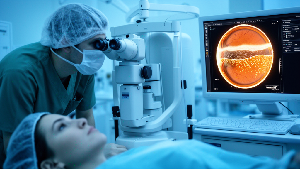
When the femtosecond laser scans the cornea to carve out the lenticule of tissue that will correct a patient’s myopia or astigmatism, the photodisruption process generates tiny pockets of gas inside the stroma. Usually these dissipate without incident, but when they accumulate into a continuous opaque layer they obscure the surgeon’s view of deeper corneal structures, making lenticule dissection harder and raising the risk of lenticule tears. Now a team at the Zhongshan Ophthalmic Center of Sun Yat-sen University in Guangzhou, China, reports that machine learning models built entirely from preoperative and surgical-planning measurements can predict both whether this complication will occur and, if it does, how extensive it will be.
The study, published in the journal Eye and Vision, was deliberately exploratory in design. The researchers retrospectively reviewed SMILE procedures performed at their center between August 2024 and August 2025, all carried out by a single experienced surgeon to eliminate inter-surgeon variability. From this pool they identified 72 eyes in which a clearly visible opaque bubble layer had formed during posterior lenticule scanning, the stage the authors singled out because an opaque bubble layer at this point carries the greatest clinical relevance. Each case eye was matched with two control eyes of the same age and sex, yielding a final dataset of 216 eyes from 216 patients, with only one eye per patient included so that inter-eye correlation was not an issue. Control eyes showed no visible opaque bubble layer or only minimal scattered bubbles that never formed a continuous opaque sheet.
The eligibility criteria were strict. Participants ranged from 18 to 45 years old, had spherical equivalents between −1.00 and −10.00 diopters with cylinder no greater than 6.00 diopters, and had never undergone prior corneal refractive or ocular surgery. Anyone with active ocular disease, including keratoconus, severe dry-eye syndrome with ocular surface damage, or uveitis, was excluded, as were eyes with poor-quality surgical videos, intraoperative complications other than an opaque bubble layer, or incomplete preclinical data. Because complete preoperative examination data were required for inclusion, the final analytical dataset contained no missing values and no imputation was needed.
Quantifying the complication was itself a technical exercise. The researchers extracted a representative single frame from each intraoperative video at the moment the posterior lenticule surface scanning was complete, the point at which the opaque bubble layer was most prominent and its boundary clearly identifiable. After converting the image to 8-bit grayscale, they applied a single uniform threshold selected during a pilot assessment and then applied consistently to all images without image-specific adjustment. The relative opaque bubble layer area, expressed as a percentage of the total posterior corneal surface area, became the primary quantitative index, and the measurements were performed by observers masked to the patients’ clinical data and model predictors.
The modeling pipeline drew on an unusually rich feature set: 54 candidate preoperative and surgical variables spanning demographics, refraction, corneal morphology measured with Pentacam HR Scheimpflug tomography, corneal optical density, intraocular pressure, and surgical-planning parameters such as cap diameter and thickness, femtosecond laser energy, and residual stromal thickness. Using the automated machine learning framework PyCaret, the team evaluated 15 classification algorithms for the binary question of whether an opaque bubble layer would occur, and 19 regression algorithms for the continuous question of how large it would be. The classification dataset of 216 eyes was split into 172 eyes for training and cross-validation and 44 eyes for internal hold-out testing, stratified by opaque bubble layer status. The regression task was restricted to the 72 opaque-bubble-layer-positive eyes, divided into 57 for training and 15 for hold-out testing, a two-stage design that avoided modeling a zero-inflated outcome distribution. Hyperparameter tuning and model selection happened exclusively within the training sets using 10-fold cross-validation.
For predicting whether an opaque bubble layer would form, the Extra Trees classifier came out on top. On the untouched internal hold-out test set of 44 eyes, which contained 15 opaque-bubble-layer-positive and 29 control eyes, it achieved an area under the receiver operating characteristic curve of 0.885 (95% confidence interval 0.871–0.899) and an accuracy of 0.820 (95% CI 0.797–0.842), with a Kappa coefficient of 0.658 indicating moderate-to-good agreement between predictions and observed outcomes. Shapley additive explanations analysis, the interpretability technique known as SHAP, then ranked the driving features. Femtosecond laser energy, intraocular pressure, residual stromal thickness, 10-millimeter corneal volume, and total astigmatism were the top five factors, all positively correlated with opaque bubble layer risk, meaning higher values corresponded to an elevated probability of intraoperative bubble formation.
The direction of some associations was less intuitive. A lower index of surface variance, a thicker corneal cap, smaller anterior surface numerical eccentricity, and smaller posterior corneal astigmatism were each associated with a reduced risk in the model, implying that corneas with regular surfaces and flatter, more spherical shapes may paradoxically be more prone to bubble-layer formation. The authors suggest a mechanical explanation: when femtosecond laser scanning occurs at a relatively shallow depth, gas bubbles may become trapped in the densely arranged collagen fibers of the anterior stroma, making dissipation difficult, while a higher intraocular pressure, which is linked to greater corneal stiffness, may further impede bubble escape.
The second stage of the framework tackled severity rather than occurrence. Among the 19 regression algorithms, random forest regression performed best on the internal hold-out test of 15 opaque-bubble-layer-positive eyes, producing a mean absolute error of 2.89% (95% CI 2.72%–3.06%) and a root mean square error of 3.37% (95% CI 3.25%–3.48%) for the relative opaque bubble layer area. Here the SHAP analysis told a different story. Age and corneal optical density within the central 2-millimeter zone of the anterior 120-micrometer corneal layer were negatively associated with area, while the keratoconus index emerged as the most influential positive predictor. The authors note this does not contradict the occurrence findings: bubble-layer formation depends chiefly on gas retention, whereas its extent depends on the capacity for bubble diffusion and coalescence. A lower optical density, they propose, reflects a looser collagen fiber arrangement in the anterior stroma, offering less structural resistance and more favorable conditions for bubbles to spread and merge.
The team is careful about what these numbers do and do not mean. Because the design was retrospective, single-center, and 1:2 matched, the predicted probabilities were not calibrated to absolute clinical risk, so no probability threshold can be recommended. Age and sex, having defined the matching procedure, were excluded from the occurrence-classification predictors, and matching selectively restricted the control sample, potentially reducing transportability to unmatched populations. The bootstrap confidence intervals quantify sampling variability of the fixed test-set predictions only and do not propagate uncertainty from preprocessing, tuning, or model selection, so the point estimates should be read descriptively. Intraoperative factors such as docking quality, suction stability, and ocular movement were never quantitatively recorded, and correlated corneal parameters may destabilize SHAP-based feature importance, which the authors frame strictly as model-derived associations rather than causal mechanisms.
Those caveats notwithstanding, the framework points toward a practical future in which a patient’s routine preoperative workup feeds a risk model before the laser ever fires. Some predictors, notably femtosecond laser energy and residual stromal thickness, are surgical-planning parameters rather than purely biometric ones, so the models may be most useful for risk assessment after an initial surgical plan has been generated. The authors explicitly caution that the findings do not justify changing laser energy settings, and that a prospective evaluation would be required before any parameter recommendations could be made. External validation and recalibration in consecutive multicenter cohorts with a natural opaque bubble layer prevalence, standardized grading, and additional corneal biomechanical measurements are the stated next steps before the approach could be considered for clinical use. Until then, the study stands as a hypothesis-generating demonstration that the seeds of an intraoperative complication may already be visible in the preoperative numbers.
Subject of Research: Machine learning prediction of opaque bubble layer formation and area during small-incision lenticule extraction
Article Title: Preoperative predictive factors for opaque bubble layer formation and area during small-incision lenticule extraction: predictive models based on machine learning
Article References: Peng, C., Chen, X., Yang, Y., Ren, H., Huang, Y., Qiu, J., Li, Y., Yu, K., & Zhuang, J. (2026). Preoperative predictive factors for opaque bubble layer formation and area during small-incision lenticule extraction: predictive models based on machine learning. Eye and Vision, 13(1), Article 39. https://doi.org/10.1186/s40662-026-00509-w
Image Credits: AI Generated
DOI: 10.1186/s40662-026-00509-w
Keywords: SMILE, opaque bubble layer, femtosecond laser, machine learning, refractive surgery, cornea, SHAP, Extra Trees, random forest, intraocular pressure, corneal tomography, risk prediction
News Source: Ophelia Keating. (October 29, 2026). Machine Learning Predicts Opaque Bubble Layer Risk During Keyhole Laser Vision Surgery. Scienmag.