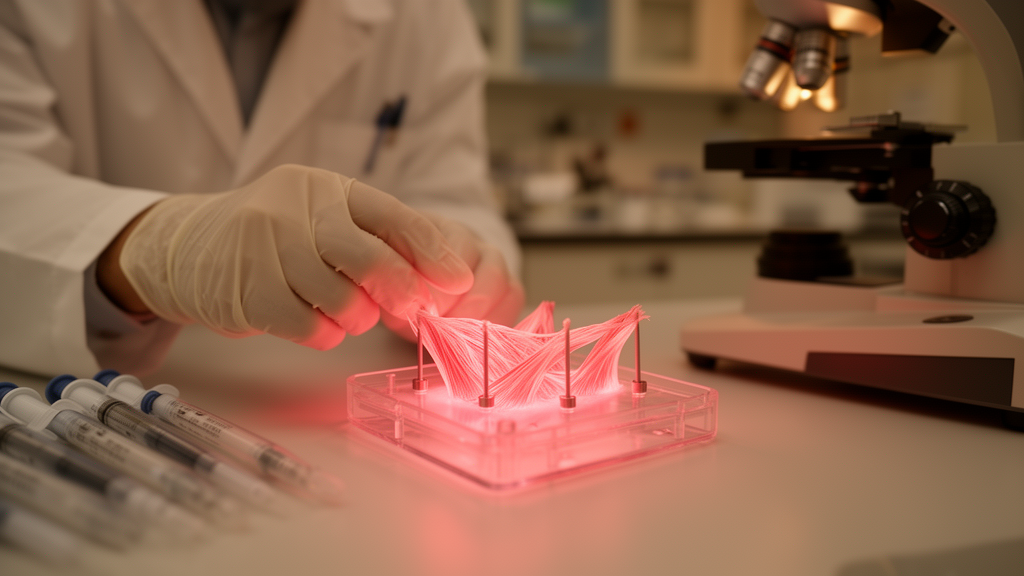
Researchers have grown functioning three-dimensional human muscle tissue from small muscle samples taken from children with rare rheumatic diseases, offering a new way to study muscle weakness in conditions that have been difficult to model in the laboratory. The proof-of-concept study, published in BMC Rheumatology, generated engineered muscle constructs known as myobundles from pediatric patients with juvenile dermatomyositis (JDM), immune-mediated necrotizing myopathy (IMNM), and inflammatory arthritis-related weakness.
Muscle weakness is a prominent feature of several rare pediatric rheumatic diseases, but the mechanisms behind the dysfunction remain poorly understood, partly because physiologically relevant laboratory models have been lacking. The new study set out to test whether meaningful muscle models could be built from the very limited tissue that clinicians can safely obtain from young patients with these conditions.
Myobundles were treated as technical replicates within each donor. Because the study included a single donor per condition, the results are presented descriptively, without inferential statistics.
Myoblast purity was high across all donors, ranging from 83 to 96 percent. The cells from the IMNM donor appeared elongated and spindle-shaped. The researchers assessed purity and morphology using desmin and vimentin co-staining, and evaluated the mature myobundles by measuring electrically evoked contractile force and twitch kinetics, alongside immunofluorescence for sarcomeric α-actinin and vimentin.
Compared with myobundles from the healthy pediatric donor, constructs from all three donors with weakness showed reduced contractility, altered striated myofiber organization, and decreased myofiber diameter.
Myobundles from the IMNM donor showed the most pronounced functional impairment, with markedly reduced force generation, slowed twitch kinetics, reduced sarcomeric α-actinin-positive area, and disrupted myofiber organization. In contrast, myobundles from the arthritis-related weakness donor exhibited faster twitch kinetics and greater fatigability than controls, even though this donor had the highest clinical strength scores of the three patients.
The findings demonstrate that functional myobundles can be generated from the limited muscle tissue available in rare pediatric rheumatic diseases, and that donor-specific functional and structural differences are measurable in vitro.
The authors caution that the study is a proof of concept with a single donor per condition. Confirmation that the observed differences represent reproducible disease-associated phenotypes will require additional donors within each condition. The article was published on 8 October 2026 as an open-access Research article in BMC Rheumatology.
Subject of Research: Patient-derived engineered muscle models for studying muscle weakness in rare pediatric rheumatic diseases
Article Title: Patient-derived myobundles as a platform for modeling muscle weakness in rare pediatric rheumatic diseases: a proof-of-concept study
Article References: Simon, A., Covert, L. T., Kandil, S., Chen, J., Dvergsten, J. A., Ardalan, K., Jones, K. A., & Truskey, G. A. (2026). Patient-derived myobundles as a platform for modeling muscle weakness in rare pediatric rheumatic diseases: a proof-of-concept study. BMC Rheumatology. https://doi.org/10.1186/s41927-026-00701-4
Image Credits: AI Generated
DOI: 10.1186/s41927-026-00701-4
Keywords: juvenile dermatomyositis, immune-mediated necrotizing myopathy, myobundles, muscle weakness, pediatric rheumatology, engineered muscle tissue, type I interferon, proof-of-concept study, myogenic progenitor cells, contractile force, BMC Rheumatology, Patient-derived
News Source: Harold Sullivan. (October 28, 2026). Lab-grown muscle bundles reveal weakness patterns in rare pediatric rheumatic diseases. Scienmag.