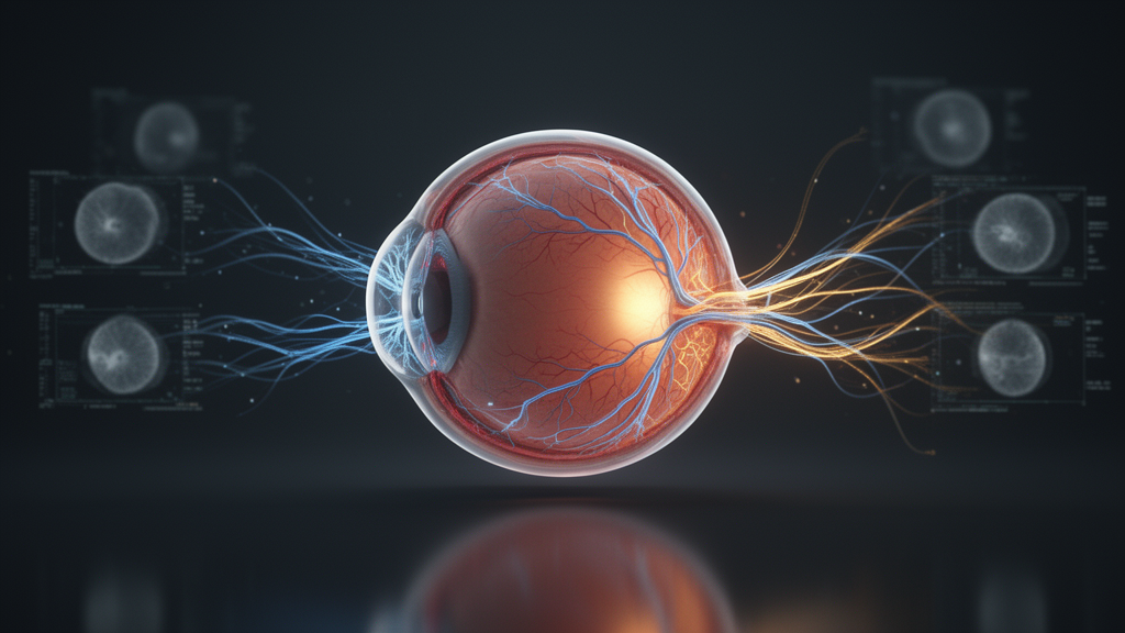
An artificial intelligence system designed to spot four major eye diseases from retinal photographs has shown just how fragile even the most impressive diagnostic models can be when they leave the laboratory. The framework, called FusionEye-Net, achieved a near-perfect 99.24 percent accuracy on its internal test set, yet when confronted with an independent collection of 4,640 fundus images gathered under different imaging conditions, its accuracy collapsed to 72.05 percent. That dramatic drop, documented in a study published in Discover Artificial Intelligence, is a vivid illustration of domain shift, the mismatch between the data a model learns from and the data it meets in the real world.
The researchers, Ali M. Duhaim and Abbas M. Al-Bakry, built their system to classify fundus photographs into four categories: cataract, diabetic retinopathy, glaucoma, and normal retina. These conditions are among the leading causes of preventable blindness worldwide, and fundus photography is a mainstay of early detection. Because trained ophthalmologists are scarce and manual review of retinal images is time-consuming, deep learning classifiers have long been proposed as a way to scale up screening. The catch, repeatedly confirmed in prior studies, is that models scoring above 95 percent internally can lose 20 to 40 percentage points when evaluated on external data from different cameras, clinics, and populations.
FusionEye-Net’s architecture is deliberately hybrid. Rather than relying on a single convolutional neural network, it runs each image through two parallel backbones and merges their outputs. EfficientNet-B3, with its Squeeze-and-Excitation attention modules, specializes in high-frequency local textures such as microaneurysms and focal lens opacities. ResNet-50, with its deep residual pathways, captures low-frequency global anatomy, including vascular configurations and optic disc boundaries. Each branch compresses its spatial feature maps through global average pooling into a vector, and the two vectors, 1,536 and 2,048 dimensions respectively, are concatenated into a unified 3,584-dimensional embedding that feeds a dense classification head ending in a four-way softmax.
Before any image reaches the network, it passes through a preprocessing pipeline designed to strip away device-specific quirks. A Hough Circle Transform isolates the circular retinal region and masks everything outside it, eliminating camera borders and vignette edges. Images are resized to 325 by 325 pixels, then contrast-limited adaptive histogram equalization is applied to the luminance channel in LAB color space with an 8-by-8 tile grid and a clipping threshold of 1.2, sharpening subtle lesions without amplifying sensor noise. Adaptive gamma correction, with exponents between 1.2 and 1.5 for dim images and below 1.0 for overexposed ones, corrects illumination inconsistencies, and a color-matching transform shifts hue distributions toward the internal training cohort’s reference mean.
The internal dataset came from a public Kaggle collection of 4,217 fundus images. A two-stage quality filter, combining automated screening for blur, saturation, exposure, and vessel visibility with a blind manual audit by a board-certified retina specialist, permanently removed 230 non-gradable samples, leaving 3,987 high-quality images. These were split 80/10/10 into training, validation, and test sets using patient-level grouping so that bilateral images from the same person never straddled partitions. Notably, the external dataset was left completely unfiltered to preserve real-world clinical noise, a deliberate choice to stress-test deployment conditions.
Training proceeded in two phases. First, with both backbones frozen, only the fusion block and classification head learned, using Adam with a learning rate of 0.001 and class-weighted losses, until validation accuracy plateaued near 94 percent. Then the final 40 layers of EfficientNet-B3 and the final 30 layers of ResNet-50 were unfrozen, with batch normalization layers kept locked, and fine-tuned at a learning rate of 0.0001 with label smoothing of 0.05. At inference, a test-time augmentation protocol averaged softmax outputs over five slightly rotated and illumination-shifted views of each image, lifting peak internal accuracy from 98.50 to 99.24 percent, with only three misclassified cases out of 396 test images and an area under the ROC curve of 1.000 for every class.
The external evaluation told a far less flattering story. On the 4,640 unfiltered external images, accuracy fell to 72.05 percent with a macro F1-score of 0.7156. The failure mode was especially dangerous for screening: the model frequently labeled diseased eyes as healthy, missing 598 diabetic retinopathy cases and 123 cataract cases by predicting normal. Diabetic retinopathy recall dropped to 0.48, while the normal class showed high recall of 0.96 but poor precision of 0.60, meaning the model defaulted uncertain cases to healthy. The authors attribute this to domain shift from different imaging devices, illumination, and patient demographics, and note it is consistent with the broader medical AI literature showing that high internal accuracy does not guarantee practical reliability.
To recover performance, the team applied a lightweight supervised domain adaptation protocol. A balanced budget of 2,000 external images, 500 per class, was set aside, split into 1,600 for fine-tuning and 400 for validation. The pre-trained weights were loaded, the same structural layers relaxed, and the network tuned for just five epochs at extra-low learning rates descending from 0.00001 to 0.000005. Crucially, these 2,000 images were removed from evaluation entirely, and final performance was measured on the remaining 2,640 locked, unseen external images. On that held-out set, accuracy reached 91.27 percent with a macro F1-score of 0.9139, cutting misclassifications by more than two-thirds. Diabetic retinopathy recall doubled from 47.72 to 90.47 percent, cataract recall rose by more than 34 percentage points, and post-adaptation AUC values ranged from 0.979 to 0.995 across classes.
Transparency was addressed through explainability analysis. Gradient-weighted Class Activation Mapping highlighted the image regions driving each prediction, and a class-specific adaptive thresholding scheme converted the heatmaps into sharp region-of-interest contours, using a lower threshold of 0.5 for diffuse conditions like cataract and diabetic retinopathy and a higher threshold of 0.6 for the localized optic disc changes of glaucoma. A retina specialist then blind-audited 200 post-adaptation feature maps, 50 per category, scoring each contour for alignment with true pathological anatomy. The resulting visual alignment index was 94.5 percent, with 189 of 200 maps judged clinically plausible. The attention patterns matched expected diagnostic reasoning: diffuse activation across the retina for cataract, vascular and macular focus for diabetic retinopathy, tight contours around the optic nerve head for glaucoma, and weak diffuse activation for normal eyes.
The study has clear limitations the authors acknowledge. The framework covers only four diagnostic categories, not the full spectrum of retinal disease. The adaptation protocol requires labeled samples from the target domain, which may not be readily available in every setting. External validation relied on a single large public dataset rather than multiple independent clinical cohorts, and the explainability audit covered a sample rather than the complete data. Even so, the central lesson stands: the team demonstrated that a modest, budget-aware adaptation effort using only 2,000 labeled external images can restore most of a model’s lost performance, and that reporting both pre- and post-adaptation external results offers a far more honest picture of clinical readiness than internal accuracy alone. Future work, the authors suggest, includes integrating vision transformers, federated learning for privacy-preserving multi-center training, multi-label classification, and uncertainty-aware predictions that flag ambiguous cases for expert review.
Subject of Research: Cross-domain deep learning classification of fundus eye diseases using hybrid CNN fusion and supervised domain adaptation
Article Title: A trustworthy cross-domain AI framework for fundus disease classification using hybrid CNN fusion and supervised domain adaptation
Article References: Duhaim, A. M., & Al-Bakry, A. M. (2026). A trustworthy cross-domain AI framework for fundus disease classification using hybrid CNN fusion and supervised domain adaptation. Discover Artificial Intelligence, 6(1), Article 967. https://doi.org/10.1007/s44163-026-02067-5
Image Credits: AI Generated
DOI: 10.1007/s44163-026-02067-5
Keywords: fundus classification, domain shift, domain adaptation, deep learning, EfficientNet-B3, ResNet-50, Grad-CAM, diabetic retinopathy, glaucoma, cataract, medical imaging AI, external validation
News Source: Blake Davidson. (October 28, 2026). Hybrid AI recovers eye-disease accuracy after real-world domain shift. Scienmag.