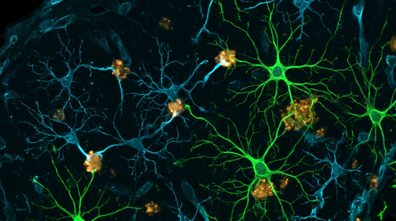
A scientific review in Frontiers in Neuroscience demonstrates that microglia and astrocytes act as a critical functional checkpoint in Parkinson’s disease, determining whether pathogenic alpha-synuclein seeds are eliminated or redistributed across the brain. Authored by Dr R. M. Meade from the University of Bath, the assessment synthesises recent cellular findings to show that non-neuronal cells are neither simple scavengers nor passive casualties. Instead, the biological outcome of glial involvement depends on a delicate balance between aggregate internalisation, lysosomal processing, and extracellular release. When degradation operates effectively, glia form an efficient sink for extracellular seeds, preventing them from inducing aggregation in neighbouring neurons. However, when degradative pathways become overwhelmed, glia can facilitate the persistence, modification, and transmission of seed-competent assemblies. This perspective reframes glial biology, positioning cellular clearance pathways as central regulators of pathological propagation.
The defining pathology of Parkinson’s disease involves the accumulation of aggregated alpha-synuclein within Lewy bodies and Lewy neurites. Misfolded assemblies act as structural templates that recruit soluble endogenous protein, driving self-propagating inclusion formation. Experimental preformed fibril models have shown that exogenous aggregates enter neurons and induce insoluble, hyperphosphorylated inclusions. In wild-type mice, injected fibrils initiate progressive pathology along anatomically connected circuits, ultimately causing dopaminergic neurodegeneration. Because microglia and astrocytes surround neuronal synapses and cell bodies, they intercept extracellular fibrils before they reach adjacent neurons. Whether this encounter restricts disease progression or drives further spread depends entirely on whether glial uptake results in complete degradation or incomplete proteolysis that releases active seeds back into the surrounding tissue. Consequently, resolving the intracellular journey of these intercepted fibrils has become a vital objective in neurodegeneration research.
As resident phagocytes of the central nervous system, microglia rapidly clear extracellular protein aggregates. Mechanistic studies in human primary and induced pluripotent stem cell-derived microglia reveal that fibril uptake occurs through MerTK, a receptor tyrosine kinase mediating homeostatic phagocytosis, with receptor inhibition markedly reducing internalisation. However, microglial clearance capacity depends heavily on inflammatory state. Exposure to pro-inflammatory interferon-gamma reduces fibril uptake and arrests phagosome maturation, demonstrating that inflammatory signaling can impair seed removal. To handle proteotoxic stress, microglia can also transfer fibrillar cargo through filamentous actin-dependent connections to neighbouring microglia for cooperative degradation, which reduces inflammatory activation and improves survival. In humanised mice, transplanted human stem cell-derived microglia restricted fibril spread from the striatum to the substantia nigra, protecting dopaminergic neurons. These in vivo findings substantiate the concept that uncompromised microglial phagocytosis provides a functional defense against anatomical disease transmission.
Astrocytes also take up extracellular fibrillar alpha-synuclein, directing it to lysosomal compartments where degradation occurs far more rapidly than in neurons. Direct comparisons indicate that preformed fibrils have a half-life of three to six hours in primary microglia and astrocytes, compared to approximately twelve hours in primary neurons. In animal models, labelled fibrils accumulate quickly in glia before clearance, whereas neuronal accumulation is slower and more persistent. Nonetheless, astrocytic clearance can fail under sustained stress. In primary cortical co-cultures, heavy uptake of oligomeric alpha-synuclein leads to incomplete lysosomal degradation, generating persistent intracellular deposits and mitochondrial abnormalities. Furthermore, genetic factors impair astrocytic function; astrocytes carrying the Parkinson’s disease-linked LRRK2 G2019S mutation show reduced capacity to internalise and clear fibrillar aggregates via endolysosomal pathways compared with wild-type cells. In astrocyte-neuron co-cultures, intact astrocytes degrade fibrils and protect neurons, but genetic vulnerability or lysosomal overload compromises this defensive buffering role.
The ultimate fate of internalised seeds depends on the endolysosomal system, yet incoming aggregates can directly disrupt this machinery. In murine BV2 cells and primary microglia, fibrillar, but not monomeric, alpha-synuclein inflicts lysosomal damage, with autophagy inhibition triggering mitochondrial dysfunction and microglial death. Recent work has revealed a direct molecular mechanism: preformed fibrils impair microglial lysosomal acidification by binding ATP6V0C, an essential subunit of the lysosomal vacuolar-type ATPase proton pump, thereby disrupting pump assembly. Restoring ATP6V0C expression improved lysosomal acidification, accelerated alpha-synuclein degradation, and reduced pathology in a mouse model. This demonstrates that fibrillar aggregates can actively disarm their own degradative clearance. When lysosomal acidification fails, the internal environment loses the enzymatic capacity required to neutralise pathogenic seeds, trapping the cell in a state of chronic organelle stress. Consequently, aggregate exposure directly compromises the cellular systems that healthy glia rely upon to restrict neuropathological seeding.
Lysosomal trafficking does not guarantee aggregate destruction, as partial enzymatic proteolysis can generate more virulent seeds. In mouse models, microglial TREM2-dependent uptake is followed by cleavage by the lysosomal protease asparagine endopeptidase. Rather than destroying the fibril, asparagine endopeptidase generates carboxy-terminally truncated fragments spanning residues 1 to 103, which exhibit enhanced seeding potency. Deleting either TREM2 or asparagine endopeptidase reduced pathology, proving that incomplete cleavage actively remodels fibrils into more pathogenic species. A similar dependence on intact degradation occurs in astrocytes. Primary astrocytes rapidly degrade fibrils and prevent neuronal seeding, but pharmacological disruption of lysosomal function reverses this protection. Astrocytes with compromised lysosomes induce neuronal pathology following fibril exposure, whereas astrocytes with intact lysosomes do not. Degradative capacity within the receiving glial cell therefore directly determines whether seed potency is extinguished or magnified. This reinforces the finding that uptake without complete proteolysis converts a protective sink into a generator of hyper-potent seeds.
When intracellular degradation fails, glia redirect persistent alpha-synuclein toward secretion into the extracellular environment. Lysosomal overload promotes the release of insoluble aggregates from fibril-loaded microglia and macrophages via an LRRK2- and Rab10-dependent extracellular vesicle pathway. In activated microglia, fibril exposure impairs autophagy through PELI1-dependent degradation of lysosomal associated membrane protein 2, driving the secretion of alpha-synuclein-containing exosomes that induce aggregation in recipient neurons. Striatal injection of exosomes from fibril-treated microglia into mice produced phosphorylated inclusions across anatomically connected regions. Heightened vesicle release also follows the acidification deficits caused by ATP6V0C disruption. These observations reveal that internalisation without destruction turns glia into propagation intermediates, repackaging seeds into vesicles that facilitate neuronal uptake and template endogenous aggregation. Thus, secretory pathways allow stressed glia to relieve their degradative burden at the direct expense of neighboring neuronal proteostasis.
In addition to vesicle release, seeds and organelles move directly between cells through intercellular physical connections. In human neuron-microglia models, alpha-synuclein-induced mitochondrial damage causes mitochondrial DNA release into the cytoplasm, activating the cGAS-STING innate immune pathway. This activation triggers the formation of tunneling nanotube-like conduits that enable directional transfer of aggregates and damaged mitochondria from neurons to microglia, where microglial lysosomes degrade the transferred organelles. Astrocytes form similar nanotube connections, transferring aggregates to adjacent astrocytes when lysosomal capacity is saturated, while receiving functional mitochondria in return to support metabolic health. Furthermore, in human co-cultures of astrocytes and microglia, astrocytes internalise more aggregates and tend to release them, whereas microglia exhibit superior degradative capacity and receive aggregates directly from astrocytes, demonstrating a cooperative clearance network. These structural networks illustrate that direct cell-to-cell transfer can either rescue overburdened cells or distribute pathological aggregates across cellular communities.
Understanding divergent findings across published studies requires recognizing that aggregated alpha-synuclein comprises structurally heterogeneous assemblies. In human stem cell-derived microglia, oligomers, preformed fibrils, and mature fibrils elicit distinct transcriptional and inflammatory responses, with mature fibrils causing strong inflammatory activation while standard fibrils induce limited acute cytokine release. Furthermore, fibrils amplified from post-mortem Parkinson’s disease brains trigger significantly stronger cytokine and glutamate responses in primary microglia than synthetic recombinant fibrils. Technical variables during fibril preparation also fundamentally affect experimental results. Sonication initially enhances seeding by fragmenting fibrils into smaller active units, but prolonged sonication dismantles the core beta-sheet structure and reduces seeding capacity. Standard protocols underscore rigorous validation of monomer preparation, aggregation conditions, storage, and fibril sizing, because differences in exposure concentration and duration directly influence whether glia successfully clear seeds or succumb to lysosomal overload. Consequently, experimental outcomes reflect not just glial capability, but the precise biochemical identity of the conformational species administered.
Variations in glial origin, activation state, and genetic background further explain conflicting experimental observations. Microglia and astrocytes do not adopt fixed protective or toxic phenotypes, but occupy dynamic transcriptional states shaped by age, tissue signals, and inflammatory context. Human genetics strongly connects glial pathways to Parkinson’s disease risk. Disease heritability is significantly enriched in microglial open chromatin, implicating loci such as P2RY12. Similarly, variants in GBA1 diminish lysosomal glucocerebrosidase activity, which has been linked to aggregate accumulation and intercellular spread in both neurons and glia. These cell-state nuances resolve apparent discrepancies between animal models. While microglial depletion reduced pathology in specific mouse fibril models, sustained colony-stimulating factor 1 receptor inhibition in a rat model depleted microglia without altering inclusion burden or nigrostriatal neurodegeneration, illustrating that glial contributions depend on the exact balance between uptake, processing, and release. Such disparities demonstrate that experimental model design and species differences exert profound influences on measured pathological outcomes.
Finally, the review stresses that seeded aggregate propagation must be distinguished from actual neurodegeneration. In preformed fibril models, inclusion burden does not correlate strictly with neuronal loss. Selective vulnerability in dopaminergic neurons involves additional factors, including cytosolic dopamine oxidation, reactive dopamine metabolites, mitochondrial dysfunction, and impaired autophagic proteostasis. Consequently, the capacity of glia to restrict seeding does not automatically confer neuroprotection. From a therapeutic standpoint, broadly suppressing or eliminating microglia could eliminate vital clearance mechanisms, whereas enhancing uptake alone risks overwhelming lysosomal degradation. Instead, precise therapeutic interventions should focus on enhancing lysosomal resilience and terminal degradation, such as restoring ATP6V0C to normalize lysosomal acidification or activating transcription factor EB to stimulate autophagic-lysosomal pathways. Resolving how glial cells process pathogenic seeds in vivo will be essential for developing treatments that arrest propagation without compromising essential homeostatic functions. Understanding these critical boundaries offers a clearer path toward targeting non-neuronal cells in Parkinson’s disease therapy.
Subject of Research: Glial mechanisms of alpha-synuclein aggregate uptake, degradation, and propagation in Parkinson's disease
Article Title: Glial handling of alpha-synuclein seeds: uptake, degradation and redistribution in pathological propagation
Article References: Meade, R. M. (2026). Glial handling of alpha-synuclein seeds: uptake, degradation and redistribution in pathological propagation. Frontiers in Neuroscience, 20, Article 2017355. https://doi.org/10.3389/fnins.2026.2017355
Image Credits: AI Generated
DOI: 10.3389/fnins.2026.2017355
Keywords: alpha-synuclein, Parkinson's disease, microglia, astrocytes, lysosomal degradation, preformed fibrils, extracellular vesicles, neurodegeneration, Glial, handling, seeds, uptake
News Source: Diana Fleming. (November 6, 2026). How Glial Cells Determine Whether Parkinson’s Aggregates Clear or Propagate. Scienmag.