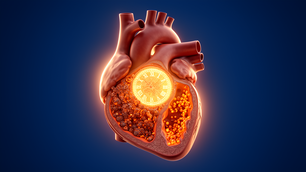
Blood pressure is not a flat number across the day. In healthy people it falls by 10 to 20 percent during sleep, dips to a nocturnal nadir, and surges in the early morning hours. That rhythmic rise and fall is not an accident of daily habits; it is orchestrated by an internal timekeeping system that coordinates the sympathetic nervous system, vascular tone, and endocrine signals across a 24-hour cycle. A new review published in Frontiers in Cardiovascular Medicine argues that when this circadian architecture breaks down, the damage may run far deeper than lost sleep. The authors, Hui Zeng, Jiawen Lin, and Jun Chen of Shanghai Yangpu Hospital of Traditional Chinese Medicine and Macau University of Science and Technology, propose that circadian disruption may set the stage for a specific form of iron-driven cell death called ferroptosis, quietly eroding the health of blood vessels and kidneys and thereby raising the risk of hypertension.
The review, published on 16 October 2026, builds on a well-established observation: blood pressure follows a pronounced circadian rhythm, yet the mechanisms by which temporal imbalance translates into sustained vascular and renal injury have remained unclear. The authors’ answer is a proposed framework they call the circadian–ferroptosis axis. In their formulation, circadian misalignment creates a temporal mismatch between the generation of oxidative stress and the body’s antioxidant defenses, lowering the threshold at which cardiovascular and renal tissues succumb to ferroptosis. This process, they suggest, may amplify hemodynamic stress, impair endothelial integrity, and disturb renal sodium handling, ultimately contributing to the development and progression of high blood pressure. Crucially, the authors are careful to frame this as an integrative and testable hypothesis rather than an established single causal pathway, noting that direct causal evidence remains limited.
To understand the proposal, it helps to start with the molecular clock itself. The mammalian circadian system is hierarchical, anchored by a central pacemaker in the suprachiasmatic nucleus of the hypothalamus, which synchronizes peripheral clocks distributed across organs through neural, hormonal, and metabolic cues. At the cellular level, the clock runs on an evolutionarily conserved transcription–translation feedback loop. The transcription factors BMAL1 and CLOCK form heterodimers that bind to E-box elements in promoter regions, activating clock-controlled genes including the Period and Cryptochrome families. The resulting PER and CRY proteins accumulate, form repressive complexes, and return to the nucleus to inhibit BMAL1-CLOCK activity, closing the loop and setting the roughly 24-hour period. Auxiliary loops involving the ROR and REV-ERB nuclear receptors, which competitively bind ROR response elements in the BMAL1 promoter with opposing effects, reinforce the oscillator’s stability and amplitude.
This molecular machinery does far more than keep time. Transcriptomic analyses have shown that more than 40 percent of protein-coding genes exhibit rhythmic expression in at least one tissue, including a substantial number involved in redox homeostasis. That time-dependent oscillation in antioxidant capacity allows cells to adapt dynamically to fluctuations in oxidative stress across the day-night cycle. Experimental work illustrates the principle vividly. In mice with skeletal muscle–specific mitochondrial respiratory uncoupling, mitochondrial dysfunction triggered an integrated stress response under circadian control, with stress-response transcriptional activity peaking during the active phase and circulating myokines such as FGF21 and GDF15 peaking in the early rest phase. Although mitochondrial dysfunction typically elevates oxidative stress and the risk of lipid peroxidation, no marked exacerbation of cell death was observed, apparently because key antioxidant enzymes such as GPX and NQO1 reached peak activity from the late active phase into the early rest phase, suppressing reactive oxygen species–mediated lipid peroxidation. When the temporal synchronization between antioxidant capacity and oxidative stress is disrupted, cells become highly susceptible to oxidative damage.
That vulnerability is where ferroptosis enters the picture. First conceptualized in 2012, ferroptosis is an iron-dependent form of regulated cell death characterized by uncontrolled lipid peroxidation. Excessive accumulation of mitochondrial reactive oxygen species triggers peroxidation of membrane phospholipids, initiating free radical chain reactions that compromise membrane integrity, and recent work suggests the initiating cascade may also arise from lysosomal iron pools, where redox-active iron accelerates phospholipid oxidation. Progressive membrane destabilization ultimately activates downstream events including NINJ1-mediated plasma membrane rupture. Cells counter these threats with multiple antioxidant systems, chief among them the glutathione peroxidase 4 pathway, which uses glutathione to detoxify lipid peroxides. The cystine/glutamate antiporter system Xc⁻, composed of SLC7A11 and SLC3A2, supplies the cystine needed for glutathione biosynthesis, while ferroptosis suppressor protein 1 acts independently of GPX4 by regenerating lipid-soluble antioxidants. Notably, recent studies show that ferroptosis can also occur spontaneously under endogenous oxidative stress: a GPX1-OSBPL8 axis at the endoplasmic reticulum mitigates ROS-induced lipid peroxidation, and loss of either component allows persistent intracellular ROS to trigger ferroptosis even without exogenous inducers.
Emerging evidence indicates that susceptibility to ferroptosis is not constant but rhythmically modulated by the clock. In aged mice, chronic circadian disruption induced DNA damage and ferroptosis in the spleen: 14-month-old Per1/Per2 double-knockout mice showed significantly increased splenic lymphocyte death, elevated malondialdehyde and ROS, upregulated transferrin receptor 1, increased pro-ferroptotic genes including Cd36, Atm, and Acsl4, and decreased anti-ferroptotic GPX4. In a separate line of work, testosterone deficiency downregulated BMAL1 and promoted hepatocellular ferroptosis, exacerbating diet-induced fatty liver disease, while aberrant upregulation of CRY2 in trophoblasts from women with recurrent miscarriage suppressed BMAL1, reduced GPX4, xCT, and SOD2 expression, and induced ferroptosis. The review’s authors argue that similar interactions may plausibly occur in vascular tissue, where endothelial cells and vascular smooth muscle cells—both metabolically active and rich in polyunsaturated membrane lipids—are particularly susceptible to ferroptotic damage, although this link remains to be directly established.
The vascular story is where the framework becomes most concrete. Peripheral molecular clocks operate in endothelial cells and vascular smooth muscle cells, and experimental studies show that smooth muscle–specific deletion of BMAL1 attenuates time-of-day–dependent vascular responsiveness to vasoconstrictors such as norepinephrine and serotonin, blunting the diurnal oscillations of vascular contractility and the synchronization of blood pressure rhythms. Under circadian disruption, persistent alterations in vascular tone and blood flow may produce abnormal shear stress patterns that generate ROS in endothelial cells, while clock-controlled antioxidant genes such as GPX4 lose their rhythmic expression and decline, compromising the GSH-GPX4 detoxifying axis. Excessive oxidative stress impairs endothelial nitric oxide synthase function and reduces nitric oxide bioavailability, favoring vasoconstriction and increased peripheral resistance. The resulting rise in blood pressure may further exacerbate endothelial injury, creating a self-amplifying vicious cycle. Hypertension and endothelial dysfunction also increase vascular permeability, allowing low-density lipoprotein to enter the arterial wall where it becomes oxidized, and oxidative stress can push vascular smooth muscle cells toward foam cell–like and osteogenic-like phenotypes that promote lipid deposition and vascular calcification.
The circadian system also shapes the inflammatory milieu. Rev-erbα directly represses Ccl2 expression in macrophages, limiting macrophage adhesion and migration, while BMAL1 regulates NRF2-dependent antioxidant responses such that its loss increases oxidative stress and enhances IL-1β production. Time-of-day control of mitochondrial function regulates NLRP3 inflammasome activation in macrophages, and myeloid-specific Bmal1 deletion enhances inflammasome activity. TNF-α secretion itself exhibits circadian variation influenced by BMAL1-dependent regulation and rhythmic TLR4 signaling, and TNF-α activates NF-κB signaling that promotes endothelial adhesion molecules including ICAM1 and VCAM1, facilitating leukocyte adhesion and vascular inflammation. The authors suggest that this inflammatory and oxidative microenvironment may increase vascular-cell susceptibility to ferroptotic injury, providing an additional potential link between circadian disruption and vascular damage, though they emphasize that these studies do not directly establish causation.
The kidney adds a second organ-level dimension. Renal function and tissue oxygenation exhibit pronounced circadian variation, and rhythmic oxygen levels can regulate HIF-1α activity and reset circadian clocks. HIF-1α can suppress ferroptosis by promoting SLC7A11 translation through YTHDF1, while hypoxia can disrupt circadian oscillations through intracellular acidification and suppression of mTORC1 activity, a pathway whose inhibition reduces GPX4 expression and promotes ferroptosis in other models. Renal NRF2 expression oscillates under clock control, and during periods of reduced NRF2 activity, exposure to contrast agents exacerbates lipid peroxidation and ferroptosis in tubular epithelial cells, whereas pharmacological NRF2 activation suppresses ferroptosis and mitigates kidney damage. The CLOCK–BMAL1 heterodimer also suppresses TGF-β–COX2 profibrotic signaling, and CLOCK deficiency abolishes the rhythmic expression of SOD1 in the kidney, increasing oxidative stress. The authors propose that ferroptotic renal injury may compromise tubular reabsorptive and excretory functions, disturbing sodium and water homeostasis and the regulation of blood pressure rhythmicity.
Preclinical evidence in hypertension models lends tentative support. Sestrin2 overexpression suppressed angiotensin II–induced ferroptosis in human umbilical vein endothelial cells, restored mitochondrial morphology, and reduced both systolic and diastolic blood pressure in mice, while renal Sestrin2 silencing exacerbated oxidative stress and raised blood pressure. Pharmacological inhibition of ferroptosis with ferrostatin-1 ameliorated angiotensin II–induced hypertension and attenuated vascular smooth muscle cell ferroptosis through the SLC7A11/GPX4 pathway. In a stress-induced hypertension model, resveratrol activated the AMPK/Sirt3 pathway in neurons of the rostral ventrolateral medulla, reducing oxidative stress, ferroptosis, and sympathetic activity, ultimately lowering blood pressure. SIRT7 alleviated renal ferroptosis and fibrosis in hypertensive mice by promoting KLF15/Nrf2 signaling, and lenvatinib-induced YAP inhibition promoted endothelial ferroptosis and hypertension that ferrostatin-1 markedly improved. Intermittent fasting, acting through the BMAL1–AMPK/NRF2 axis, has also been shown to attenuate ferroptosis by restoring antioxidant defenses.
Yet the clinical picture remains open. No clinical studies have directly evaluated ferroptosis-targeted interventions for blood pressure reduction or restoration of circadian blood pressure patterns, and validated biomarkers that specifically and dynamically reflect ferroptosis in hypertensive patients are lacking, since markers such as malondialdehyde, 4-hydroxynonenal, GPX4, SLC7A11, and ACSL4 also arise from broader oxidative and inflammatory processes. The authors propose integrating multimodal ferroptosis-related signatures with 24-hour ambulatory blood pressure monitoring to test whether time-dependent changes in ferroptotic signals track abnormal blood pressure rhythms. Meanwhile, behavioral and chronotherapeutic approaches already show promise: hypertensive patients who maintained a fixed bedtime for two weeks showed significant reductions in nocturnal blood pressure, bedtime dosing of olmesartan plus amlodipine reduced nocturnal systolic pressure without increasing nocturnal hypotension risk, and timed sodium intake may matter as much as total salt. The authors envision constructing a 24-hour ferroptosis susceptibility landscape through time-series multi-omics and real-time redox imaging, targeting ferroptosis at optimal circadian phases to maximize efficacy while minimizing off-target effects. For now, the circadian–ferroptosis axis remains a hypothesis—but one that could reshape how hypertension is prevented, measured, and treated.
Subject of Research: The proposed circadian–ferroptosis axis linking circadian rhythm disruption, oxidative stress, and ferroptotic cell death to vascular and renal injury in hypertension
Article Title: Circadian rhythm dysregulation and ferroptosis in hypertension: mechanisms and therapeutic implications
Article References: Zeng, H., Lin, J., & Chen, J. (2026). Circadian rhythm dysregulation and ferroptosis in hypertension: mechanisms and therapeutic implications. Frontiers in Cardiovascular Medicine, 13, Article 1889670. https://doi.org/10.3389/fcvm.2026.1889670
Image Credits: AI Generated
DOI: 10.3389/fcvm.2026.1889670
Keywords: hypertension, circadian rhythm, ferroptosis, oxidative stress, endothelial dysfunction, BMAL1, GPX4, kidney, vascular remodeling, chronotherapy, blood pressure, sleep disruption
News Source: Ophelia Keating. (October 17, 2026). How Broken Body Clocks and Iron-Driven Cell Death May Fuel Hypertension. Scienmag.