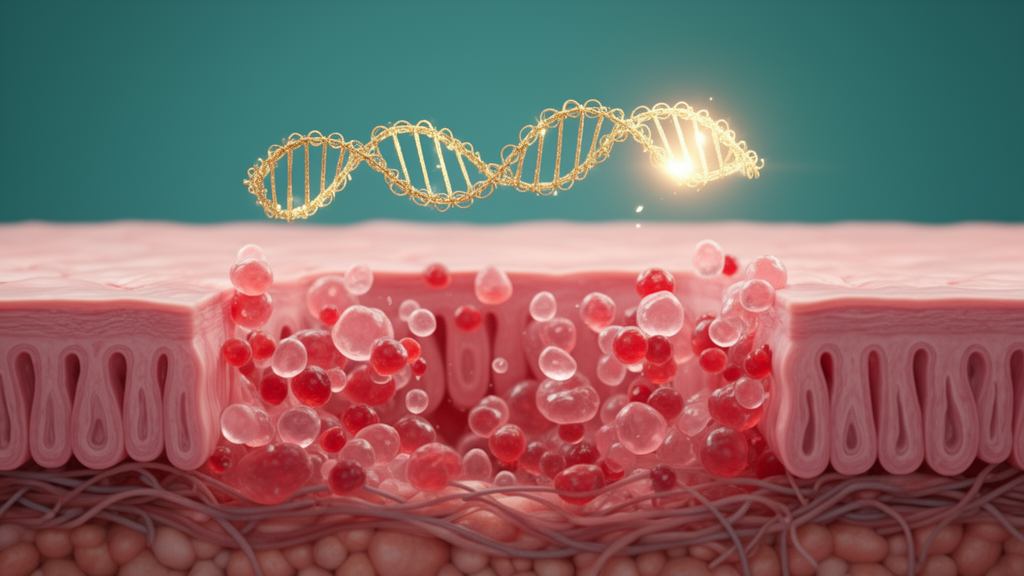
Scientists have uncovered a molecular trick that colorectal tumors use to fuel their own spread: a shortened RNA splice variant of the CALD1 gene, produced at abnormally high levels in cancer tissue, appears to help tumor cells migrate and invade distant organs. The finding, published in Molecular Cancer, traces a complete regulatory chain from a splicing protein called hnRNPA1, through the tumor-specific CALD1 variant dubbed CALD1S, to the motor protein MYH9, and links that chain to advanced disease stage and poorer survival in patients with colorectal cancer.
Alternative splicing is one of the genome’s most powerful sources of biological diversity. After a gene is transcribed into messenger RNA, cellular machinery can cut and rejoin the transcript in different patterns, so a single gene can yield many distinct protein products. In healthy tissues this process is tightly controlled, but in cancer the controls often slip. Tumors frequently produce splice variants that normal cells never make, and some of these variants endow cancer cells with new abilities, including the capacity to break away from the primary tumor and colonize other tissues. Despite growing recognition of this phenomenon, the functional relevance of individual splice isoforms in colorectal cancer, and the splicing regulators that direct their generation, have remained poorly charted.
To map that landscape, the research team, led by Jin Wang, Hulin Ma and senior author Yong Peng of West China Hospital, Sichuan University, together with colleagues at several Sichuan University institutes, performed RNA sequencing on five paired samples of colorectal tumor tissue and adjacent normal tissue. The comparison revealed widespread splicing aberrations across the cancer transcriptome: 1,048 significantly altered splicing events distinguished tumor from normal tissue. Among the affected transcripts, one stood out. A short isoform of the CALD1 gene, which encodes caldesmon, a well-known regulator of the actin cytoskeleton, was missing two specific segments, exons 3b and 4, and was preferentially expressed in tumor tissue. The team named this truncated version CALD1S.
The researchers verified the presence of CALD1S using several independent techniques, including reverse transcription PCR, quantitative RT-PCR and immunoblotting, confirming that the variant is both transcribed and translated in cancer cells. When they examined clinical samples, the pattern was striking: elevated CALD1S expression correlated significantly with advanced tumor stage, with lymph node metastasis, and with poorer patient survival. In other words, the more CALD1S a tumor produced, the more aggressive the disease appeared to be.
Functional experiments then tested what the variant actually does. Using gain- and loss-of-function assays in laboratory-grown colorectal cancer cells, the team found that CALD1S promoted cell migration and invasion, the two cellular behaviors that underpin metastasis. Notably, the variant exerted negligible impact on cell proliferation, indicating that its role is specifically in dissemination rather than in tumor growth. In animal models, forcing CALD1S expression enhanced liver metastatic dissemination, one of the most feared outcomes of colorectal cancer, while removing the variant impaired the cells’ ability to spread.
The next question was how the variant is generated in the first place. Splicing decisions are made by RNA-binding proteins that recognize sequence cues on pre-messenger RNA. Through RNA immunoprecipitation and minigene splicing assays, the researchers identified hnRNPA1, a heterogeneous nuclear ribonucleoprotein with established roles in RNA processing, as the splicing factor that binds the CALD1 pre-mRNA and promotes skipping of exons 3b and 4. When hnRNPA1 was abundant, the cell produced more CALD1S; when the team depleted hnRNPA1, CALD1S production fell and the cancer cells lost much of their migratory and invasive capacity. Critically, restoring CALD1S expression partially rescued those impaired phenotypes, demonstrating that CALD1S is a functional downstream effector of hnRNPA1 rather than an incidental byproduct.
To understand how CALD1S exerts its pro-metastatic effect at the protein level, the researchers turned to co-immunoprecipitation and mass spectrometry-based proteomics, methods that reveal which proteins physically associate with one another inside cells. These approaches identified MYH9, the heavy chain of non-muscle myosin IIA, as a CALD1S-interacting protein. MYH9 is a molecular motor that generates contractile forces in the actin cytoskeleton, and its involvement provides a plausible mechanical explanation for the variant’s effect: by engaging the myosin machinery, CALD1S may reorganize the cytoskeletal forces that cancer cells use to squeeze through tissue barriers. Knocking down MYH9 attenuated the CALD1S-driven cell migration, supporting that interpretation.
The clinical data reinforced the mechanistic story. In lymph node-positive colorectal cancer tumors, CALD1 and MYH9 protein levels exhibited a strong positive correlation, consistent with the two proteins acting together in the same pathway. Moreover, patients whose tumors showed higher combined CALD1 and MYH9 expression had an unfavorable prognosis. The study therefore delineates what the authors describe as an hnRNPA1-CALD1S-MYH9 regulatory axis that couples aberrant alternative splicing to metastatic progression in colorectal cancer.
The implications reach in two directions. As a biomarker, CALD1S offers a potential way to identify patients whose tumors are primed for spread, information that could guide surveillance intensity and treatment decisions, particularly since the variant was preferentially expressed in tumor tissue rather than normal tissue. As a therapeutic target, the axis suggests several intervention points, from blocking hnRNPA1’s binding to the CALD1 pre-mRNA to disrupting the CALD1S-MYH9 interaction, although the authors note that hnRNPA1 participates in many RNA-processing events, so selective targeting will require care. The work was funded by the Natural Science Foundation of Sichuan and the 1.3.5 Project for Disciplines of Excellence at West China Hospital, and the open-access article was published on 17 October 2026 after peer review.
For a disease that remains one of the leading causes of cancer death worldwide, largely because of metastasis to the liver and other organs, the identification of a tumor-specific splice variant with a defined upstream driver and a defined downstream partner is a meaningful step forward. It adds colorectal cancer to the growing list of malignancies in which the splicing code itself, not just the genes it encodes, becomes a driver of disease, and it suggests that reading that code carefully could yield both better prognostic tools and new points of attack.
Subject of Research: Cancer Biology
Article Title: Tumor-associated CALD1 splicing variant driven by hnRNPA1 promotes colorectal cancer metastasis
Article References: Wang, J., Ma, H., Liu, W., Wu, Q., Lu, Y., Ming, Y., Guo, J., Li, J., Dai, L., Wang, T.-H., Yang, J., & Peng, Y. (2026). Tumor-associated CALD1 splicing variant driven by hnRNPA1 promotes colorectal cancer metastasis. Molecular Cancer. https://doi.org/10.1186/s12943-026-02812-5
Image Credits: AI Generated
DOI: 10.1186/s12943-026-02812-5
Keywords: colorectal cancer, alternative splicing, CALD1, hnRNPA1, MYH9, metastasis, Molecular Cancer, biomarker, Tumor-associated, splicing, variant, driven
News Source: Nathaniel Bowman. (October 17, 2026). Hidden RNA splice variant drives colorectal cancer spread, study finds. Scienmag.