Phosphorylation of the histone variant H2AX, driven by a regulatory axis involving the SET oncoprotein and protein phosphatase 2A, maintains the stemness and tumorigenic capacity of glioma stem cells, according to a peer-reviewed laboratory study published in Genes & Diseases. Researchers from The Ohio State University and Columbus Academy identified that H2AX and its Ser139-phosphorylated form, known as γH2AX, are significantly enriched in glioma stem cell populations compared to their differentiated counterparts, playing an essential functional role in sustaining tumor-initiating capabilities.
Glioblastoma represents an aggressive primary brain tumor characterized by pronounced resistance to standard therapeutics and high rates of recurrence. A major factor contributing to this clinical challenge is the presence of glioma stem cells, a dedicated subpopulation of tumor-initiating cells that drive tumor progression and post-treatment relapse. While existing cancer literature has implicated specific transcription factors and broader chromatin remodeling complexes in preserving stem-like states, the precise contributions of individual histone variants to glioma stem cell maintenance have remained largely unresolved.
To investigate epigenetic factors regulating these cells, the research team conducted comparative proteomics analysis evaluating glioma stem cells alongside corresponding differentiated glioma cells. In their initial model system, GBM387 cells were maintained under suspension spheroid culture conditions to preserve their intrinsic stemness characteristics, or alternatively cultured in the presence of 10% fetal bovine serum to induce cellular differentiation. Subsequent proteomic profiling specifically focused on histone family members, generating volcano plot analyses that revealed differential expression patterns between the two cellular states.
Validation experiments confirmed that both total H2AX and Ser139-phosphorylated γH2AX were consistently elevated in glioma stem cells relative to differentiated cells across multiple glioblastoma models. Immunoblotting demonstrated this pattern across three independent lines of glioblastoma xenograft-derived stem cells compared against cells differentiated using either fetal bovine serum or bone morphogenetic protein 4. To substantiate these cellular states, researchers assessed a panel of stemness genes, confirming that elevated histone expression tracked closely with established stemness signatures.
The elevated presence of H2AX and γH2AX was further evaluated across established glioblastoma cell lines, including U251, T98G, and LN229. These lines were initially maintained in adherent serum conditions to foster non-stem populations, transferred into suspension cultures utilizing Neurobasal medium supplemented with B27, epidermal growth factor, and basic fibroblast growth factor to enrich for stem-like spheres, and subsequently returned to serum-containing adherent cultures. In parallel, cell sorting based on the stem cell marker CD133 in GBM387 and U251 models demonstrated that CD133-positive subpopulations expressed higher quantified levels of both H2AX and γH2AX immediately following isolation than CD133-negative fractions.
To determine whether H2AX was functionally required to maintain stem properties, the investigators performed CRISPR-Cas9-mediated deletion of the H2AX gene. In vitro functional assays revealed that while general cellular proliferation rates remained unaffected by the deletion, the loss of H2AX significantly impaired key stem-associated traits. Specifically, H2AX-knockout cells displayed marked reductions in self-renewal capacity, diminished neurosphere formation efficiency, and impaired in vivo tumor initiation.
The consequences of H2AX depletion were subsequently tested in vivo using intracranial GBM387 xenograft models. Mice bearing intracranial tumors derived from H2AX-knockout cells demonstrated substantially prolonged overall survival compared to control counterparts, directly linking the presence of the histone variant to aggressive tumor formation within the brain environment.
Underlying transcriptional mechanisms were evaluated using RNA-sequencing and targeted expression profiling following H2AX depletion. The resulting transcriptional landscape showed significant downregulation of critical stemness- and neurodevelopment-associated genes, including CXCL12, KLF4, JAK3, and SALL4. Furthermore, the molecular profiling indicated suppression of the PI3K–AKT signaling cascade, an established pathway vital for glioma stem cell survival and self-renewal.
To establish whether Ser139 phosphorylation was the key driver of these phenotypes, the authors carried out genetic rescue experiments in H2AX-knockout cells using either wild-type H2AX or a phosphorylation-deficient S139A mutant. Reintroduction of wild-type H2AX successfully restored neurosphere formation, self-renewal, and tumor-initiating capacity. In contrast, the S139A mutant failed to rescue these properties, demonstrating that phosphorylation at the Ser139 residue is functionally indispensable for preserving the glioma stem cell compartment.
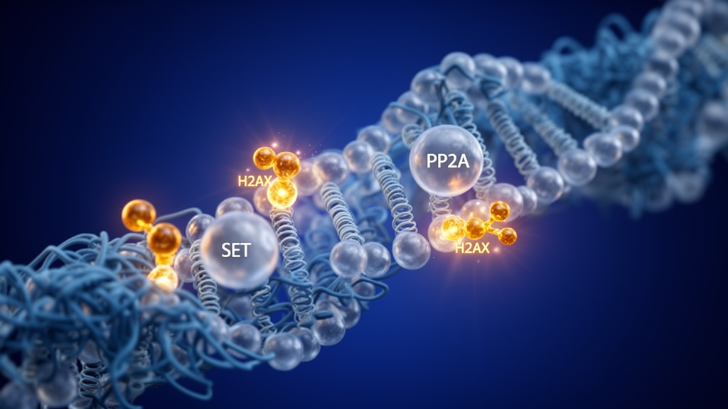
Mechanistic assays demonstrated that the elevated accumulation of γH2AX in glioma stem cells was not accompanied by elevated DNA double-strand breaks or an activated DNA damage response. Instead, proteomic and biochemical investigations revealed increased expression of the SET oncoprotein, an endogenous inhibitor of protein phosphatase 2A, which normally acts to dephosphorylate γH2AX. Knockdown of SET or pharmacological reactivation of protein phosphatase 2A using the small molecule DT-061 effectively decreased γH2AX levels, establishing a SET–PP2A–γH2AX regulatory axis that may offer a targeted therapeutic vulnerability to limit glioblastoma recurrence.
The investigation, conducted by researchers at The Ohio State University and Columbus Academy including Chunhua Han, Na Li, Ananya Banerjee, Qi-En Wang, and Arnab Chakravarti, provides extensive experimental characterization of how histone variant regulation intersects with glioma biology. A critical observation from their CRISPR-Cas9 gene editing experiments is the functional dissociation between general cell division and stemness preservation. Although deleting H2AX stripped the glioma stem cells of their hallmark self-renewal capabilities, neurosphere formation capacity, and in vivo tumorigenicity, the genetic deletion left overall cell proliferation completely intact. This distinction indicates that H2AX does not simply act as a general housekeeping factor necessary for basic cellular replication, but instead plays a selective regulatory role in preserving the specialized stem-like phenotype responsible for glioblastoma propagation.
To rigorously define the expression dynamics of H2AX and γH2AX during the transition between stemness and differentiation, the team evaluated multiple differentiation protocols across three distinct glioblastoma xenograft-derived lines. Differentiation was driven by exposing stem-like cells to ten percent fetal bovine serum or, alternatively, by treating cultures with bone morphogenetic protein 4. Across both differentiation modalities, immunoblotting revealed a pronounced reduction in H2AX and γH2AX abundance when glioma stem cells transitioned into differentiated glioma cells. To confirm that these expression shifts reflected authentic lineage changes rather than culture artifacts, the authors tested a comprehensive panel of stemness-associated marker genes across all cell populations, confirming that the decline in histone variant abundance directly mirrored the loss of verified stemness characteristics.
Complementary culturing strategies in the established glioblastoma cell lines U251, T98G, and LN229 provided further biochemical confirmation. When non-stem populations maintained in adherent serum conditions were transferred into suspension cultures utilizing Neurobasal medium enriched with B27 supplement, epidermal growth factor, and basic fibroblast growth factor, they formed floating spheroids enriched for stem cell traits. Returning these spheres to adherent conditions with serum prompted their differentiation back into non-stem glioma cells, with immunoblotting tracking the reversible upregulation and downregulation of H2AX and γH2AX. Furthermore, fluorescence-activated sorting of CD133-positive and CD133-negative subpopulations from GBM387 and U251 cells enabled immediate comparative analysis. Direct densitometric scanning of the resulting immunoblot bands demonstrated that CD133-positive stem-enriched fractions harbored substantially higher quantified relative intensity values for both H2AX and γH2AX than their CD133-negative counterparts.
The genome-wide downstream consequences of H2AX loss were comprehensively mapped through RNA-sequencing and targeted molecular validation. The resulting transcriptional profiles demonstrated that depleting H2AX triggered widespread reprogramming of gene networks essential for maintaining primitive cellular identities. Among the most significantly downregulated transcripts were CXCL12, KLF4, JAK3, and SALL4, each of which has recognized functions in neurodevelopmental programs and stemness maintenance. In parallel with the downregulation of these specific developmental regulators, H2AX-depleted cells exhibited pronounced suppression of PI3K–AKT signaling. Because the PI3K–AKT cascade represents a central signaling nexus required to sustain glioma stem cell survival and self-renewal, its disruption provides a clear molecular rationale for the impaired clonogenic growth and reduced tumorigenic potency observed in animal models.
The molecular dependency on H2AX was resolved to a single amino acid modification through rigorous rescue experiments comparing wild-type protein against a phosphorylation-deficient mutant. Introducing wild-type H2AX into knockout glioma stem cells fully rescued their neurosphere formation capacity, self-renewal potential, and tumor-initiating ability, whereas expressing the S139A mutant in which serine 139 was mutated to alanine failed to restore these malignant phenotypes. Crucially, while γH2AX is canonical in the scientific literature as a classic surrogate marker for DNA damage, the authors demonstrated that its accumulation in glioma stem cells occurred in the complete absence of elevated DNA double-strand breaks or heightened DNA damage checkpoint activation. This uncoupling established that γH2AX in glioblastoma functions as an active epigenetic regulator of the stem cell state rather than merely reflecting genomic instability or persistent genotoxic stress.
The discovery of the SET–PP2A–γH2AX axis provides an actionable biochemical explanation for how glioblastoma stem cells sustain this phosphorylated state. Quantitative proteomic and biochemical screening identified high levels of the SET oncoprotein, a recognized endogenous inhibitor of protein phosphatase 2A, in glioma stem cells. By inhibiting protein phosphatase 2A, elevated SET prevents the enzymatic dephosphorylation of γH2AX, resulting in sustained histone phosphorylation. Experimental knockdown of SET or chemical activation of protein phosphatase 2A with the small-molecule activator DT-061 effectively decreased γH2AX abundance. In vivo intracranial xenograft models reinforced these mechanistic insights, as mice bearing H2AX-knockout tumors demonstrated substantially prolonged survival compared to controls, directly linking histone phosphorylation to lethal tumor progression. While these findings highlight targeting the SET–PP2A regulatory circuit as a viable strategy to deplete the glioma stem cell pool and limit post-surgical tumor recurrence, the authors explicitly emphasize that the detailed downstream mechanisms through which the PP2A–γH2AX axis sustains stemness warrant continued investigation in future preclinical studies.
Subject of Research: Regulation of glioma stem cell maintenance by SET-PP2A-driven H2AX phosphorylation
Article Title: H2AX phosphorylation sustains glioma stem cell properties
Article References: H2AX phosphorylation sustains glioma stem cell properties. Original publication (Bibliographic details unavailable.)
Image Credits: AI Generated
DOI: Not provided
Keywords: glioblastoma, glioma stem cells, H2AX, γH2AX, SET oncoprotein, protein phosphatase 2A, PP2A, DT-061, GBM387, CD133, PI3K-AKT signaling, CXCL12, KLF4, JAK3, SALL4, neurosphere formation, intracranial xenografts, CRISPR-Cas9 deletion
News Source: Nathaniel Bowman. (November 20, 2026). H2AX Phosphorylation Driven by SET and PP2A Sustains Glioma Stem Cells. Scienmag.