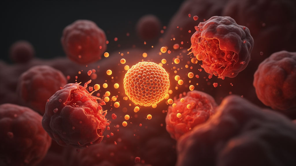
A calcium peroxide core wrapped in a copper-doped zeolite framework and studded with glucose oxidase has shown in mice that it can shrink tumors by starving cancer cells of sugar while simultaneously igniting two forms of programmed cell death, pyroptosis and cuproptosis, that together rally the immune system.
The design, called CaO2/CZ/GOX, is a cascade: each component feeds the next. Calcium peroxide nanoparticles donate hydrogen peroxide and calcium ions. The copper-doped ZIF-8 metal-organic framework releases copper ions in the acidic tumor environment. Glucose oxidase consumes glucose, the fuel tumor cells burn voraciously, and in doing so generates more hydrogen peroxide. That peroxide then feeds Fenton-like reactions driven by copper, producing hydroxyl radicals and other reactive oxygen species. The result is a self-amplifying storm of oxidative stress concentrated inside mitochondria, the energy factories of the cell.
The characterization work behind this construct was thorough. Transmission electron microscopy showed that bare calcium peroxide particles form irregular, rough agglomerates, but once confined within the copper-ZIF-8 scaffold they become uniform, well-dispersed nanoparticles with smooth edges. Dynamic light scattering confirmed a gradual increase in hydrated particle size with a polydispersity index consistently below 0.2, indicating a stable, even distribution. X-ray diffraction retained the characteristic peaks of both calcium peroxide and the ZIF-8 framework, while X-ray photoelectron spectroscopy identified copper in the divalent state and calcium ions, confirming successful loading of both components.
Glucose oxidase loading was verified by SDS-PAGE, which showed a protein band matching free GOX, and by BCA protein assay, which measured a loading efficiency of 4.3 percent. Inductively coupled plasma analysis found the particles contained 6.14 percent calcium and 2.17 percent copper by mass. Critically, the encapsulated enzyme kept working: after 24 hours in serum-containing buffer, the nanoparticles consumed glucose at rates comparable to free glucose oxidase, while control particles without the enzyme showed negligible depletion. The particles also remained stable, with no aggregation in water, buffered saline, or 10 percent fetal bovine serum over seven days.
Release behavior matched the chemistry of the tumor microenvironment. At pH 5.0, mimicking the acidity around tumors, 75 percent of the glucose oxidase was released within 24 hours, far exceeding release at the neutral pH 7.4 of healthy tissue. Calcium ion release was likewise efficient under acidic conditions. Methylene blue decolorization assays showed that the full nanosystem degraded the dye most efficiently at pH 5.0 in a concentration-dependent manner, confirming that the enzyme-supplied peroxide was fueling copper-mediated radical generation. Both glucose consumption and hydrogen peroxide production rose with glucose concentration, demonstrating substrate-dependent catalytic activity.
In Hepa1-6 mouse liver cancer cells, the nanoparticles were taken up progressively over four hours and proved potently cytotoxic, while showing negligible toxicity toward AML12 normal hepatocytes even at high concentrations. The selectivity appears metabolic: cancer cells with vigorous glucose metabolism activate the enzyme and, combined with the acidic exterior, trigger framework dissociation and release of calcium ions, reactive oxygen species, and copper. Normal cells with low glycolytic flux and a neutral environment cannot easily ignite the cascade. Antitumor efficacy climbed stepwise from calcium peroxide alone, through copper-ZIF-8 and the combined framework, to the complete three-component system.
The cell-death mechanism was traced in detail. Fluorescent probes showed that calcium and copper ions accumulated selectively in mitochondria, disrupting ion homeostasis and collapsing mitochondrial membrane potential, as revealed by JC-1 staining. Immunofluorescence and western blotting showed reduced levels of FDX1 and LIAS, proteins central to copper-induced cell death, alongside elevated oligomerized DLAT, the aggregation signature of cuproptosis. This copper-driven proteotoxic stress converged with oxidative damage on the Caspase-1/GSDMD pathway: cleaved caspase-1 and the pore-forming GSDMD-N fragment reached their highest levels in the full nanosystem group. Microscopy captured the morphological hallmarks of pyroptosis, cellular swelling with bubble-like protrusions and membrane rupture, and the cells released the highest levels of IL-1β and lactate dehydrogenase among all treatment groups.
That inflammatory death translated into immune signaling. The treated cells exposed calreticulin on their surfaces and released HMGB1 from their nuclei, the classic damage-associated molecular patterns of immunogenic cell death that flag dying tumor cells for phagocytosis and activate CD8-positive T cells. In tumor-bearing mice, flow cytometry revealed significantly increased CD8-positive and CD4-positive T cells within tumors, elevated dendritic cell maturation markers CD80 and CD86 in both spleen and tumor, and raised serum levels of the inflammatory cytokines IL-6 and TNF-α. Fluorescence imaging showed the nanoparticles accumulated preferentially in tumors, peaking at 12 hours and persisting to 72 hours while organ signals faded, a distribution attributed to the enhanced permeability and retention effect.
Safety and durability were also addressed. Intravenous dosing produced no pathological changes on histological examination of major organs, with blood counts and liver and kidney function markers remaining in normal ranges through 30 days, and hemolysis stayed below 5 percent even at 150 micrograms per milliliter. In a rechallenge experiment, cured mice inoculated with fresh cancer cells on the opposite flank showed almost no recurrent tumor growth, and their spleens contained markedly increased numbers of CD44-positive, CD62L-negative effector memory CD8-positive T cells, indicating that the treatment had established long-term immune memory against the cancer.
The authors caution, in effect, that this remains early preclinical work: the efficacy and immune findings come from a single mouse liver cancer cell line with small groups of five animals, and the in vitro mechanistic studies, however detailed, were performed in cultured cells. Translating a glucose-hungry, copper-loaded, pyroptosis-triggering nanoparticle into human cancer therapy will require far more testing. Still, the study offers a concrete demonstration that metabolic starvation, mitochondrial disruption, and immunogenic cell death can be engineered into one self-reinforcing nanoscale package, providing experimental evidence for combination strategies in tumor immunotherapy.
Subject of Research: A cascade nanoplatform combining glucose oxidase, calcium peroxide, and copper-doped ZIF-8 that induces pyroptosis and cuproptosis to activate antitumor immunity in a mouse liver cancer model.
Article Title: Multifunctional cascade nanosystem consumes glucose to trigger enhanced pyroptosis and cuproptosis for tumor immunotherapy
Article References: Dong, C., Zhu, L., Liu, H., Jin, S., Wang, Q., Teng, D., Sun, Z., Liu, B., Zhang, H., Dong, P., Du, J., Sui, G., & Wang, H. (2026). Multifunctional cascade nanosystem consumes glucose to trigger enhanced pyroptosis and cuproptosis for tumor immunotherapy. Materials Today Bio, 41, Article 103772. https://doi.org/10.1016/j.mtbio.2026.103772
Image Credits: AI Generated
DOI: 10.1016/j.mtbio.2026.103772
Keywords: nanomedicine, pyroptosis, cuproptosis, tumor immunotherapy, glucose oxidase, calcium peroxide, metal-organic framework, immunogenic cell death, mitochondrial stress, hepatocellular carcinoma, reactive oxygen species, preclinical study
News Source: Nathaniel Bowman. (October 29, 2026). Glucose-Eating Nanoparticle Fires Up Two Cell-Death Pathways to Fight Tumors. Scienmag.