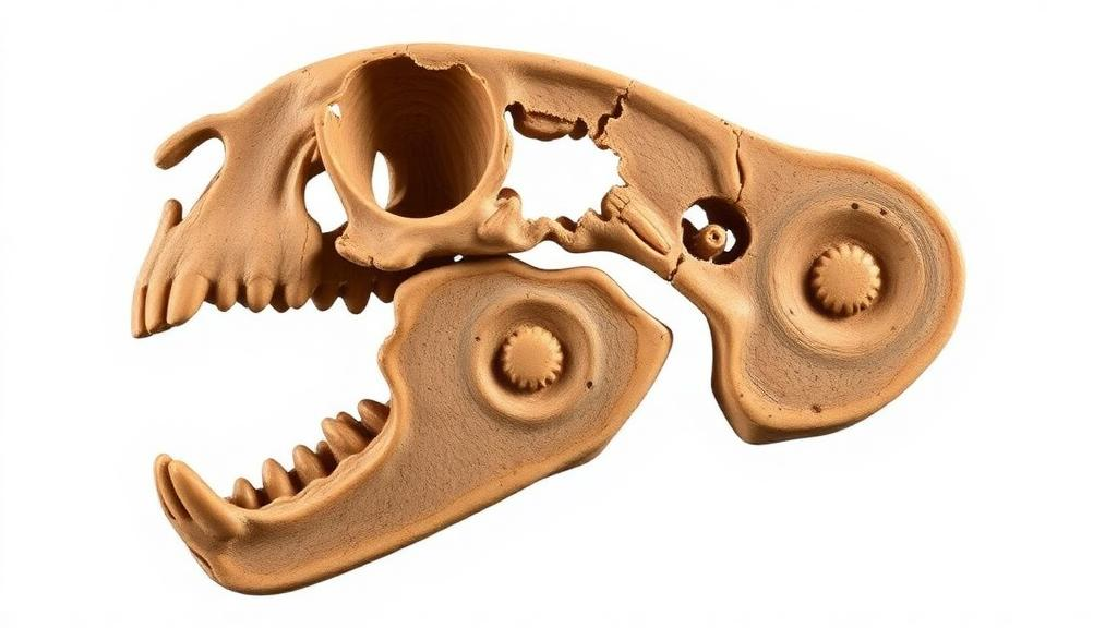
A tiny fossil mammal from the Early Cretaceous of China has given paleontologists their clearest view yet of how the mammalian middle ear broke free from the jaw. In a study published in Developmental Biology Advances, researchers describe the middle ear region of Cokotherium jiufotangensis, an early eutherian that lived roughly 120 million years ago. Using new micro-CT scans of the holotype specimen, the team virtually reconstructed the ectotympanic, a ring-shaped bone that anchors the eardrum, and combined this fossil evidence with developmental data from living mice to reconstruct the full evolutionary history of a structure most readers have never heard of: the Meckelian cartilage.
The Meckelian cartilage is a rod of cartilage that runs along the inner side of the lower jaw in jawed vertebrates. In most vertebrates it remains cartilaginous, but in early mammaliaforms, the forerunners of modern mammals, it was uniquely capable of turning into bone, leaving a bony element housed within a groove on the medial surface of the mandible. Developmental and fossil evidence shows that this structure plays a transient but crucial role in forming both the mandible and the middle ear. In extant mammals, its anterior portion contributes to the mandibular symphysis at the chin, while its posterior portion ossifies to form the malleus and incus, the middle ear bones that are homologous with the articular and quadrate bones of non-mammalian vertebrates.
In mice, the developmental sequence is remarkably precise. The cartilage elongates rapidly at embryonic days 14 to 15, forming the major cartilaginous element of the mandibular region, while dentary ossification begins at embryonic day 13. Degradation of the intermediate portion starts at embryonic day 16, and the anterior and posterior portions disconnect at embryonic days 18 to 19. At birth, the middle ear remains connected to the mandible through the cartilage. The anterior connection to the dentary disappears at postnatal days 3 to 4, and the posterior separation from the malleus follows at around postnatal day 7, leaving only a small residual nodule that forms the anterior ligament of the malleus and the sphenomandibular ligament.
The fossil record mirrors this ontogeny. Across early mammaliaforms, the arrangement of the Meckelian cartilage, ectotympanic, and malleus evolved through three recognizable stages: the Mandibular Middle Ear of the earliest diverging mammaliaforms such as morganucodontans, in which the ear bones sit in a trough on the jaw; the Transitional Middle Ear of stem clades such as eutriconodontans and symmetrodontans, in which the ear bones have shifted medially but remain linked to the jaw by an elongate ossified cartilage; and the Detached Middle Ear characteristic of early therians and all living mammals. The morphology of the Meckelian groove, ectotympanic, and malleus therefore serves as a key indicator of the evolutionary stage of the middle ear in any given fossil taxon.
Cokotherium occupies a pivotal position in this sequence. Earlier work had reported a vestigial ossified Meckelian cartilage in this species, but the original study did not recover middle ear bones from the skull region, obscuring any assessment of whether the cartilage was still connected to the ear. In the new analysis, the team scanned the holotype specimen, IVPP V23387, at a resolution of 15.138 micrometers per pixel and reconstructed the ectotympanic virtually from incomplete and slightly compressed fragments preserved between the two mandibles. The right ectotympanic is nearly complete and shows a hook-like outline with an anterior and a posterior crus, and notably no styliform process, the anterior projection that in early mammaliaforms represents a vestigial remnant of the ancestral three-pronged ectotympanic.
The absence of the styliform process matters because it indicates that the anterior limb of the ectotympanic, which in more primitive mammaliaforms articulated with the Meckelian cartilage, had been completely lost. Although the malleus itself is not preserved in the specimen, the authors argue that the complete loss of the anterior limb reinforces the interpretation that the Meckelian cartilage was no longer connected to the middle ear. The ossified cartilage in Cokotherium is extremely gracile and confined to the posterior part of the mandible near the root of the angular process, a morphology the authors interpret as functionally independent from other bony elements. This makes Cokotherium the first early therian in which both the ossified Meckelian cartilage and the ectotympanic are known, offering the first direct evidence for evaluating the jaw-ear connection in early therians.
The study places this finding within a broader pattern of parallel evolution. Middle ear detachment evolved independently multiple times among major mammaliaform clades, including australosphenidans, multituberculates, haramiyidans, and therians. The earliest documented detachment occurred in Late Jurassic haramiyidans and multituberculates, predating similar transformations in other clades by roughly 40 million years. In those groups, the Meckelian groove is commonly absent, and the accelerated reduction of the cartilage has been hypothesized as an adaptation linked to palinal chewing. Strikingly, the ectotympanic in Late Jurassic haramiyidans retains a plesiomorphic three-pronged outline, showing that the Meckelian cartilage can be lost independently of ectotympanic shape, a reflection of developmental plasticity. In cladotherians, by contrast, gradual transformations of both the cartilage and the ectotympanic suggest a different pathway, hypothesized to be driven primarily by selection for more sensitive hearing, particularly at high frequencies.
Integrating the fossil and developmental evidence, the authors propose two evolutionary trajectories for the reduction of the Meckelian cartilage in early mammaliaforms, distinguished by different configurations of the cartilage and the middle ear bones. Three major transformations stand out: the cartilage ossified and its intermediate portion was reduced at the origin of mammaliaforms; it shifted medially in transitional clades; and it became vestigial and posteriorly confined before disappearing entirely. The sequence parallels the fate of the cartilage in mouse development, from the elongate embryonic rod resembling Sinoconodon, through the divided groove of juvenile Morganucodon resembling the embryonic disconnection stage, to the vestigial remnant of Cokotherium, which the authors compare to the residual nodule seen in mice at postnatal day 7, after the ear has detached at postnatal day 4.
One intriguing discrepancy emerges from this comparison. In living mammals, the anterior release, the breakdown between the cartilage and the mandible, occurs before the posterior release between the cartilage and the ear. The fossil evidence from Cokotherium suggests the opposite order in evolution: the posterior detachment between the ossified cartilage and the middle ear apparently occurred earlier than the anterior one. The authors propose that in early therians the ossified cartilage may still have acted as a temporary stabilizer, mechanically supporting the relatively large middle ear bones before complete detachment, whereas in extant mammals the mechanical and formative functions of the cartilage are confined to very early embryonic stages and are soon replaced by adjacent dermal bones.
The study also addresses why the cartilage ossified in the first place, something never seen in non-mammaliaform cynodonts. The earliest occurrence of an ossified Meckelian cartilage, in Early Triassic to Early Jurassic mammaliaforms, coincides with a marked miniaturization of the mandible, a process previously identified as a key driver in the origin of the mammalian squamosal-dentary jaw joint. The authors hypothesize that ossification temporarily bridged the functional gap between the middle ear and the mandible, providing mechanical stability for the reduced postdentary bones during their co-option into the ear. Its eventual loss in therians, they suggest, was driven by selective pressure for increased auditory sensitivity, particularly high-frequency hearing reflected in a micro-type middle ear and a more coiled cochlear canal, which rendered the stabilizing function obsolete. The gracile, posteriorly confined cartilage of Cokotherium thus documents the latest known stage in the gradual detachment of the mammalian hearing apparatus from the jaw, a transitional architecture that bridged the ancient jaw-based hearing system and the fully detached middle ear of modern mammals.
Subject of Research: Evolutionary transformation of the ossified Meckelian cartilage and middle ear detachment in early mammaliaforms
Article Title: Evolutionary transformations of the ossified Meckelian cartilage in mammaliaforms
Article References: Wang, H., Anthwal, N., Wang, S., & Wang, Y.-Q. (2026). Evolutionary transformations of the ossified Meckelian cartilage in mammaliaforms. Developmental Biology Advances, 17(1), Article 11. https://doi.org/10.1186/s13227-026-00265-x
Image Credits: AI Generated
DOI: 10.1186/s13227-026-00265-x
Keywords: Meckelian cartilage, middle ear evolution, Cokotherium, mammaliaforms, ectotympanic, Early Cretaceous, eutherians, paleontology, evolutionary development, mandible, fossil micro-CT, therians
News Source: Gavin Prescott. (October 29, 2026). Fossil Reveals Final Step in Mammals’ Middle Ear Detachment from Jaw. Scienmag.