Researchers published a study in the journal Cell Division on October 2, 2025, detailing how Fibroblast Growth Factor 4 (FGF4) drives the progression of triple-negative breast cancer (TNBC). The investigation focused on the mechanism by which FGF4 modulates macrophage polarization through the Interleukin 6/Signal Transducer and Activator of Transcription 3 (IL6/STAT3) signaling axis. This process creates an immune-suppressive tumor microenvironment that facilitates tumor growth and immune evasion. The study integrates bioinformatics analysis with in vitro and in vivo experimental models to provide a comprehensive view of this molecular pathway.
Triple-negative breast cancer is a particularly aggressive subtype of breast cancer characterized by the lack of estrogen receptor, progesterone receptor, and Human Epidermal Growth Factor Receptor 2 (HER2) expression. This specific molecular profile renders TNBC resistant to conventional endocrine therapies and HER2-targeted treatments, contributing to a poorer clinical prognosis. Tumors in TNBC patients are often more aggressive, prone to metastasis, and less responsive to chemotherapy and radiation, leading to higher recurrence rates. Consequently, developing new therapeutic strategies that enhance immune responses is crucial for improving patient survival and treatment outcomes.
The research team, led by Xuanhe Zhang and colleagues, utilized transcriptomic datasets from The Cancer Genome Atlas (TCGA) and the Gene Expression Omnibus (GEO) to identify FGF4-associated pathways. They employed differential gene expression analysis, Weighted Gene Co-expression Network Analysis (WGCNA), and immune infiltration profiling via the Cell-type Identification By Estimating Relative Subsets Of RNA Transcripts (CIBERSORT) algorithm. Functional annotations using Gene Ontology (GO) and Kyoto Encyclopedia of Genes and Genomes (KEGG) highlighted the IL6/STAT3 pathway as a key regulatory mechanism in TNBC samples.
In the bioinformatics phase, the analysis identified 680 differentially expressed genes between TNBC and normal samples, with 382 genes upregulated and 298 downregulated. By intersecting these differentially expressed genes with key gene modules from the WGCNA, the researchers isolated 107 candidate genes. Further network analysis using the STRING database and Cytoscape software identified four core genes: HIST1H3C, SOX2, CDH2, and FGF4. CIBERSORT analysis revealed a significant positive correlation between FGF4 expression and the infiltration of M2 macrophages, as well as a notable positive correlation with M2 macrophage-associated genes IL6 and STAT3.
To validate these computational findings, the study established FGF4-overexpressing and knockdown TNBC cell lines using MDA-MB-231 and BT-549 cells. These cells were co-cultured with THP-1 macrophages, which were differentiated using phorbol 12-myristate 13-acetate (PMA). Western blotting and quantitative reverse transcription-polymerase chain reaction (qRT-PCR) confirmed that FGF4 overexpression significantly increased the expression of IL6 and phosphorylated STAT3 in macrophages. Conversely, FGF4 knockdown led to a notable reduction in these signaling molecules, indicating that FGF4 influences macrophage function by upregulating the IL6/STAT3 signaling pathway.
Immunofluorescence staining and flow cytometry were used to assess macrophage polarization markers in the co-culture systems. In the FGF4 overexpression group, the expression of M2 markers, specifically Arg1 and CD206, was significantly increased, while M1 markers such as iNOS and CD86 were markedly reduced. In contrast, the FGF4 knockdown group showed upregulated M1 markers and downregulated M2 markers. These results support the hypothesis that FGF4 promotes M2 macrophage polarization via the IL6/STAT3 axis, shifting the immune balance toward an immunosuppressive state.
The functional consequences of this polarization were assessed by measuring the secretion of immunosuppressive cytokines and the impact on T cell activity. Enzyme-linked immunosorbent assay (ELISA) results showed that macrophages from the FGF4 overexpression group secreted significantly higher levels of IL-10 and TGF-β. T cell proliferation inhibition assays using carboxyfluorescein succinimidyl ester (CFSE) staining revealed that macrophages in the FGF4 overexpression group significantly inhibited T cell proliferation. This inhibitory effect was substantially reduced in the FGF4 knockdown group, suggesting that FGF4 enhances the immunosuppressive function of macrophages via the IL6/STAT3 axis.
Furthermore, the study evaluated the impact of M2 macrophage-derived secretions on breast cancer cell behavior. Cell counting Kit-8 (CCK-8) assays demonstrated that breast cancer cell proliferation rates significantly increased when treated with M2-polarized macrophage-conditioned medium. Transwell migration and Matrigel invasion assays showed that this conditioned medium significantly enhanced the migration and invasion of breast cancer cells. These data indicate that FGF4 drives M2 macrophage polarization through the IL6/STAT3 axis, which in turn promotes tumor cell proliferation and invasion in the breast cancer microenvironment.
In vivo experiments were conducted using an orthotopic tumor model in immunocompetent BALB/c mice. FGF4-overexpressing, knockdown, and control TNBC cells were implanted into the mammary fat pads. After 28 days, tumors in the FGF4 overexpression group were significantly heavier than those in the control group, while tumors in the FGF4 knockdown group were markedly lighter. Immunohistochemistry analysis of tumor tissues revealed that M2 macrophage markers were significantly increased in the FGF4 overexpression group, whereas M1 markers were reduced. Flow cytometry of tumor-infiltrating T cells showed that the proportions of T cells positive for functional markers such as IFN-γ, TNF-α, and Granzyme B were significantly reduced in the FGF4 overexpression group.
The study also investigated the reversibility of these effects by intervening in the IL6/STAT3 pathway. When an IL6 neutralizing antibody or a STAT3 activator was added to the FGF4 knockdown co-culture system, the expression of M2 polarization markers increased, and M1 markers decreased. This suggests that FGF4 promotes IL6/STAT3 activation, thereby driving macrophage polarization towards the M2 phenotype. The findings confirm that knocking down FGF4 or inhibiting the IL6/STAT3 signaling pathway suppresses M2 polarization and enhances immune killing, while IL6 or STAT3 activators can reverse the inhibitory effects of FGF4 knockdown.
The authors acknowledge several limitations in the study. The use of independent short hairpin RNAs (shRNAs) targeting different regions would further confirm the specificity of knockdown effects by minimizing off-target artifacts. Additionally, the exploration of additional downstream targets of the IL6/STAT3 axis remains insufficient, and further research is needed to fully elucidate the complete mechanisms of this signaling pathway. The study employed human-derived TNBC and macrophage cell lines for in vitro experiments and a murine TNBC model for in vivo studies, but the lack of further validation of FGF4 expression and its clinical prognosis in human tumor samples limits the immediate clinical applicability of the findings.
Despite these limitations, the study provides new molecular insights into the initiation and progression of TNBC. FGF4 emerges as a potential biomarker and therapeutic target, offering a theoretical foundation for developing precision immunotherapy strategies. Modulation of the IL6/STAT3 signaling axis could represent a promising intervention pathway to improve TNBC prognosis and therapeutic outcomes. Future clinical trials will be crucial in validating the effectiveness of targeting FGF4 to overcome current immunotherapy limitations in triple-negative breast cancer.
Subject of Research: The role of FGF4 in triple-negative breast cancer progression via IL6/STAT3-mediated macrophage M2 polarization and immune suppression.
Article Title: FGF4 drives tumor progression in triple-negative breast cancer via IL6/STAT3-mediated macrophage M2 polarization and immune suppression
Article References: Zhang, X., Zhang, S., & Han, Y. (2025). FGF4 drives tumor progression in triple-negative breast cancer via IL6/STAT3-mediated macrophage M2 polarization and immune suppression. Cell Division, 20(1), Article 22. https://doi.org/10.1186/s13008-025-00164-y
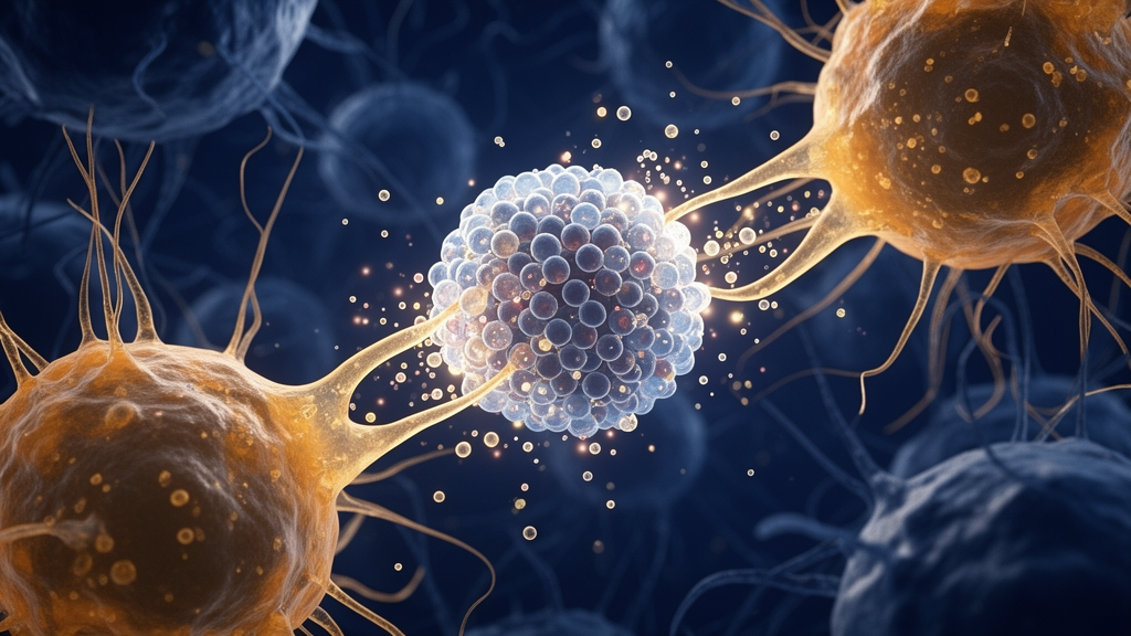
Image Credits: AI Generated
DOI: 10.1186/s13008-025-00164-y
Keywords: Triple-negative breast cancer, FGF4, Macrophage polarization, IL6/STAT3 signaling, Immune suppression, Tumor microenvironment, Cell Division, Cancer immunology, drives, tumor, progression, triple-negative
News Source: Nathaniel Bowman. (November 9, 2026). FGF4 Drives Breast Cancer via Macrophage Reprogramming. Scienmag.