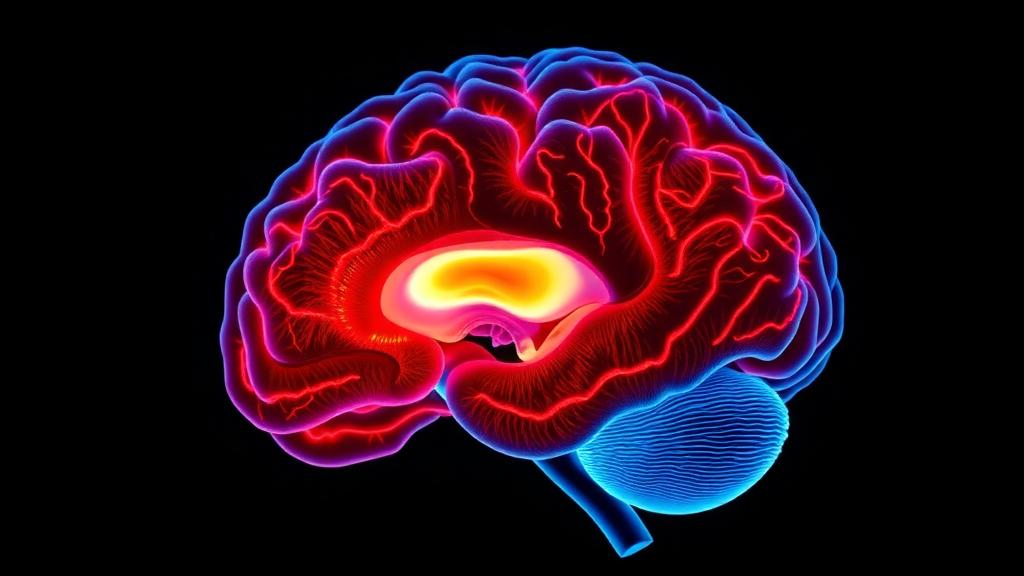
Deep inside the fluid-filled cavities of the brain sits a small, feathery structure that most people have never heard of, yet it quietly manufactures the cerebrospinal fluid that bathes and cleanses the entire central nervous system. Now, a team of researchers in China has produced some of the most direct evidence yet that this structure, the choroid plexus, becomes enlarged and inflamed in cerebral small vessel disease, a common and devastating condition that is a leading cause of stroke and cognitive decline in older adults. The findings, published in Annals of Clinical and Translational Neurology, suggest that the choroid plexus may be far more than a passive fluid factory: it could be an active driver of the inflammatory processes that damage the aging brain.
Cerebral small vessel disease, often abbreviated CSVD, refers to a spectrum of disorders affecting the brain’s tiniest blood vessels. Its fingerprints on magnetic resonance imaging are familiar to any neuroradiologist: bright patches of white matter hyperintensity, small cavities called lacunes, enlarged fluid-filled perivascular spaces, tiny cerebral microbleeds, and gradual shrinkage of brain tissue. Together, these markers underlie a substantial share of strokes and vascular cognitive impairment worldwide. Yet despite decades of study, the mechanisms that link failing small vessels to progressive brain injury remain incompletely understood, and few studies have examined how the brain’s waste-clearance systems, including the glymphatic pathway, contribute to the disease.
The choroid plexus sits at the heart of that question. This vascularized tissue, located in the lateral, third, and fourth ventricles of the brain, produces the bulk of cerebrospinal fluid and forms a critical barrier, the blood-cerebrospinal fluid barrier, that gates the passage of cells and molecules between the blood and the brain’s fluid compartments. The plexus is densely populated with immune cells and expresses a wide repertoire of inflammatory mediators, making it a key staging ground for neuroinflammation. Enlargement of the choroid plexus has previously been documented in neurodegenerative and neuroinflammatory conditions, including Alzheimer’s disease, and age-related changes in cerebrospinal fluid dynamics have been tied to structural remodeling of the tissue. Because small vessel disease and Alzheimer’s pathology so frequently coexist in elderly brains, the researchers reasoned that the choroid plexus might represent a previously overlooked biomarker in CSVD.
To test that idea, the team prospectively recruited 111 patients with cerebral small vessel disease and 69 healthy controls between 2016 and 2021 at two Chinese hospitals, with ethical approval and written informed consent. All participants underwent high-resolution brain MRI on a 3.0 Tesla scanner, including three-dimensional T1-weighted imaging, T2-weighted imaging, fluid-attenuated inversion recovery, and susceptibility-weighted sequences. Two experienced neuroradiologists, blinded to clinical information, visually rated the conventional imaging markers of small vessel disease according to the STRIVE guidelines and combined them into a total CSVD score ranging from zero to six. Meanwhile, two trained physicians independently segmented the choroid plexus volume in each ventricle using ITK-SNAP software, normalizing the measurements to total intracranial volume. The segmentation agreement was excellent, with an intraclass correlation coefficient of 0.895.
The results were striking. Patients with small vessel disease showed significantly larger choroid plexus volumes in all three ventricular compartments compared with healthy controls. In the main cohort, the median lateral ventricle choroid plexus volume was 1.74 in patients versus 1.51 in controls, the third ventricle value was 0.34 versus 0.20, and the fourth ventricle value was 0.17 versus 0.12, with all differences highly significant even after adjusting for age and sex. Crucially, the enlargement tracked with disease severity. Larger plexus volumes correlated positively with white matter hyperintensity scores, lacunes, enlarged perivascular spaces in the basal ganglia, cerebral microbleeds, and the total CSVD score, and they also correlated with overall brain atrophy. The pattern points to a widespread process affecting the entire blood-cerebrospinal fluid barrier system rather than a localized abnormality confined to a single ventricle.
The study’s most novel contribution, however, came from a subgroup of 22 patients and 10 age- and sex-matched controls who underwent a specialized imaging technique rarely applied to this disease. The researchers infused ultrasmall superparamagnetic particles of iron oxide, known as USPIO, using the clinically approved iron supplement ferumoxytol at a dose of 3 milligrams per kilogram. These nanoparticles are engulfed by activated macrophages and microglia, the immune cells that congregate at sites of inflammation, so their accumulation reveals inflammatory activity in living tissue. Because the particles are cleared from plasma within roughly 24 to 48 hours, scanning 48 hours after infusion minimizes confounding from blood retention and perfusion effects, allowing the signal changes to primarily reflect iron uptake by activated myeloid cells. The technique has proven its worth in multiple sclerosis and stroke research, but its application to the choroid plexus in small vessel disease had remained largely unexplored.
Forty-eight hours after infusion, the patients’ choroid plexus lit up. Quantitative analysis of the change in signal intensity ratio, calculated relative to neck muscle tissue before and after injection, showed significantly greater USPIO uptake in the choroid plexus of all three ventricles in patients compared with controls. The inflammation was not confined to the plexus: patients also exhibited significantly increased uptake in the hippocampus, thalamus, and medulla oblongata, revealing a spreading inflammatory process that reaches deep gray matter structures essential for cognition. Moreover, the degree of choroid plexus inflammation correlated with conventional disease markers, with lateral ventricle plexus uptake correlating with white matter hyperintensity scores and fourth ventricle uptake correlating with both white matter hyperintensities and lacunes. Uptake in the putamen and thalamus also tracked with white matter damage, suggesting that plexus inflammation participates in the broader neuroinflammatory cascade accompanying small vessel injury.
Perhaps the most conceptually important finding was the direct link between structure and function. The volume of the lateral ventricle choroid plexus correlated positively with its own inflammatory signal, and even more strongly with the inflammatory signal of the fourth ventricle plexus, relationships that persisted after adjusting for age and sex. Inflammation in the lateral ventricle plexus was also strongly correlated with uptake in the hippocampus and thalamus, hinting at a blood-cerebrospinal fluid barrier mechanism that could connect small vessel disease to Alzheimer-type neurodegeneration. The authors propose a plausible biological sequence: chronic hypoperfusion or endothelial dysfunction in small vessel disease triggers an inflammatory response in the highly vascularized plexus, prompting immune cell infiltration and cellular hypertrophy that enlarge the tissue. The inflamed plexus, in turn, may release pro-inflammatory cytokines, alter cerebrospinal fluid composition, and disrupt the barrier, reducing the efficiency of glymphatic waste clearance and creating a vicious cycle of inflammation and brain injury.
The implications extend beyond mechanism. Choroid plexus volume could serve as a practical imaging biomarker that complements existing measures of small vessel disease burden, and USPIO-enhanced MRI offers a noninvasive way to monitor neuroinflammation and evaluate the efficacy of future anti-inflammatory treatments. Interventions aimed at the plexus-cerebrospinal fluid system might eventually emerge as therapeutic strategies. The researchers caution, however, that their study is cross-sectional and therefore cannot establish causation, that the USPIO subgroup was small and requires validation in larger cohorts, and that histological confirmation of the imaging signal is still lacking. They also note that the unweighted summation of imaging markers, while clinically practical, may obscure differences in pathological importance among individual features.
Even with those caveats, the work marks a significant step forward in reframing cerebral small vessel disease as a condition in which the brain’s immune interface plays a central role. If future longitudinal studies confirm that choroid plexus enlargement and inflammation precede the appearance of white matter damage and other conventional markers, the humble fluid-producing tissue tucked inside the ventricles could become an early warning system for one of the most common threats to the aging brain, opening a window for intervention long before strokes and dementia take their toll.
Subject of Research: Choroid plexus enlargement and neuroinflammation in cerebral small vessel disease measured with conventional and USPIO-enhanced MRI
Article Title: Choroid Plexus Enlargement and USPIO‐Based Inflammatory Feature in Cerebral Small Vessel Disease
Article References: Qu, Y., Ren, X., Fan, C., Li, C., Zhang, L., Zhang, Z., Li, S., Lai, Z., Cai, B., Fu, Y., Lin, Y., & Zhu, Z. (2026). Choroid Plexus Enlargement and USPIO ‐Based Inflammatory Feature in Cerebral Small Vessel Disease. Annals of Clinical and Translational Neurology, 13(10), 2078-2087. https://doi.org/10.1002/acn3.70382
Image Credits: AI Generated
DOI: 10.1002/acn3.70382
Keywords: cerebral small vessel disease, choroid plexus, USPIO, neuroinflammation, MRI, white matter hyperintensities, glymphatic system, blood-cerebrospinal fluid barrier, ferumoxytol, brain atrophy, microglia, cognitive decline
News Source: Cassandra Pierce. (October 6, 2026). Enlarged Choroid Plexus Reveals Hidden Brain Inflammation in Small Vessel Disease. Scienmag.