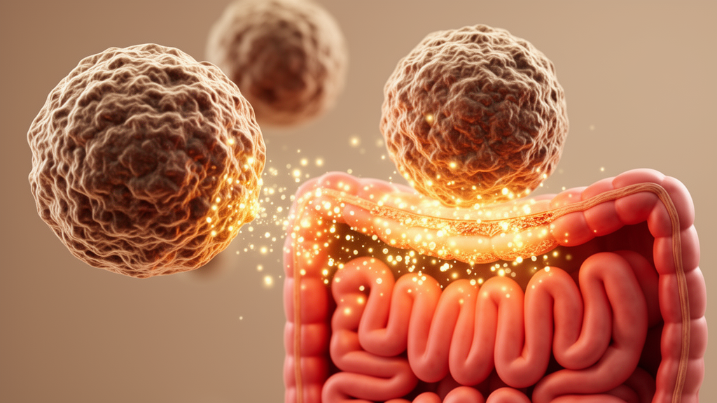
Engineered macrophages that secrete the anti-inflammatory signaling molecule interleukin-10 (IL-10) alleviated disease symptoms in a mouse model of ulcerative colitis, according to a new preclinical study. The cell therapy, administered intraperitoneally at the onset of dextran sulfate sodium (DSS) exposure, stabilized body weight, reduced diarrhea and rectal bleeding, and largely prevented the colon shortening that characterizes the model. The findings, reported in a study of DSS-induced colitis in mice, also revealed time-dependent shifts in the immune landscape of the inflamed gut and changes in the composition of the gut microbiota, offering a detailed picture of how the treatment may operate.
The rationale for the approach stems from a long-standing puzzle in inflammatory bowel disease research. IL-10 is a pleiotropic cytokine with potent anti-inflammatory properties and acts as a master regulator of intestinal immune balance, suppressing pro-inflammatory cytokines and chemokines while curbing the activation of macrophages and naïve CD4+ T cells. The importance of this pathway is underscored by animal genetics: mice lacking IL-10 or its receptor develop severe spontaneous colitis, and human neonates with defective IL-10 signaling develop severe enterocolitis shortly after birth. Yet despite encouraging results in preclinical models of inflammatory bowel disease, clinical trials of recombinant human IL-10 in patients produced disappointing outcomes.
Researchers attribute those failures largely to the limitations of systemic drug delivery. Injected IL-10 fails to reach effective concentrations within intestinal tissues, and its immunosuppressive effects can be counteracted by concurrent immunostimulatory actions elsewhere in the body. This has pushed investigators toward strategies that deliver the cytokine locally to inflamed gut tissue. Macrophages, central components of the innate immune system and pivotal guardians of intestinal homeostasis, presented an attractive vehicle. In inflammatory bowel disease, particular macrophage populations can themselves drive inflammatory responses and tissue damage, but their plasticity means their behavior can potentially be redirected for therapeutic benefit.
In the new study, the team used RAW264.7 macrophages, a cell line derived from BALB/c mice, engineered via lentiviral transduction to stably secrete IL-10. As a control, they generated ConM cells carrying an empty vector. Both cell types were injected intraperitoneally into C57BL/6 mice at the start of a ten-day DSS regimen, which induces colitis by damaging the epithelial lining of the colon. The untreated model group, receiving only phosphate-buffered saline, developed the expected constellation of disease: marked weight loss, diarrhea, hematochezia, and a disease activity index that climbed steadily over the treatment period.
The IL-10-secreting cells substantially blunted this decline. Treated mice maintained more stable body weight, produced more formed stool, and recorded lower disease activity scores than their untreated counterparts. Colon length, a standard proxy for inflammation-driven shortening, told a striking story: the model group’s colons averaged 5.35 ± 0.32 centimeters, while IL-10M-treated animals measured 7.42 ± 0.47 centimeters, statistically indistinguishable from the healthy control group at 7.67 ± 0.39 centimeters. Control macrophages carrying the empty vector partially ameliorated colon shortening, reaching 6.25 ± 0.47 centimeters, but failed to relieve weight loss or rectal bleeding. Post hoc comparisons between the two cell types confirmed that IL-10 secretion was responsible for the superior efficacy across the major endpoints.
Histological examination of colon tissue sections reinforced the clinical picture, with IL-10M-treated mice showing significantly lower histological injury scores than the model group. Yet a curious discrepancy emerged when the researchers measured cytokine levels directly. At the terminal time point on day 10, IL-10 concentrations in whole-colon homogenates showed only a non-significant increase in the treated group, even as the pro-inflammatory mediators IL-1β and IL-6 were significantly suppressed. The authors suggest this may reflect the timing of sampling: flow cytometry showed that GFP-labeled macrophages had largely cleared from the colon by the late phase of observation, meaning locally secreted IL-10 may have acted earlier or in a cell-proximal manner that bulk tissue ELISA at a single terminal time point could not fully capture.
That flow cytometry data also revealed something important about how the therapy reshapes the immune environment. Rather than simply converting inflammatory macrophages into anti-inflammatory ones in a fixed, binary fashion, the treatment appeared to modulate host macrophage phenotypes in a time-dependent manner. At day 3, IL-10M treatment increased the proportion of endogenous GFP-negative macrophages expressing CD206, a marker associated with tissue-reparative and anti-inflammatory activity, and raised the overall CD206 index. By day 6, the population of GFP-negative macrophages expressing CD86, a marker linked to inflammatory activation, showed a decreasing trend. This pattern suggests a dynamic, phased reprogramming of the intestinal immune landscape rather than a single-step switch.
The study also documented substantial changes in the gut microbiota of treated animals. Metagenomic profiling revealed that IL-10M treatment was associated with increased abundance of several bacterial taxa, including Bacteroides acidifaciens, Muribaculum intestinale, and a species designated D. freteri, alongside reduced levels of M. schaedleri, compared with untreated DSS-exposed mice. These shifts are biologically plausible given the close interconnection between intestinal inflammation, epithelial barrier integrity, and microbial ecology. However, when the researchers compared IL-10M directly against ConM, only the reduction in M. schaedleri reached statistical significance; the increases in the other taxa did not. This indicates that the macrophage vehicle itself may account for a substantial portion of the observed microbial remodeling, with sustained IL-10 expression contributing modest or additive effects rather than acting as the sole determinant.
The authors are careful to note that the study cannot establish whether these microbiota changes directly contributed to disease improvement or occurred secondarily as a consequence of reduced inflammation and restored barrier function. Testing that causal relationship, they suggest, will require future experiments involving fecal microbiota transplantation, antibiotic depletion followed by recolonization, or the use of germ-free mice. Similarly, the allogeneic nature of the cell therapy—RAW264.7 cells originate from BALB/c mice while recipients were C57BL/6—may have facilitated immune clearance of the administered macrophages, which could limit long-term persistence and uncontrolled expansion but also complicates interpretation of the durability of benefit.
Several other limitations frame the findings. The study relied on a single chemically induced model of colitis rather than models that more closely recapitulate the chronic, relapsing nature of human ulcerative colitis, and all experiments were conducted in mice, leaving open the question of whether comparable effects would emerge in human tissue. The IL-10 measurement discrepancy, the transient presence of the engineered cells, and the modest differential microbiota effects relative to the control macrophages all underscore that the mechanism of action is likely multifactorial. Nevertheless, the results establish that IL-10-secreting macrophages can outperform unmodified macrophages in this model, achieving near-complete protection of colon length and symptom control where the control cells fell short. The work adds to a growing body of research exploring cell-based delivery platforms for cytokine therapy, and the authors suggest that future studies should focus on extending these observations to additional models and ultimately evaluating whether the approach can be translated toward human inflammatory bowel disease.
Subject of Research: IL-10-secreting engineered macrophage cell therapy for DSS-induced colitis in mice
Article Title: IL‐10‐secreting engineered macrophages alleviate DSS‐induced colitis with macrophage phenotypic modulation and gut microbiota remodeling in mice
Article References: Liu, H., Liu, J., Peng, S., Zhao, J., Yang, F., Zhang, L., Wang, S., Zhang, L., & Sun, G. (2026). IL ‐10‐secreting engineered macrophages alleviate DSS ‐induced colitis with macrophage phenotypic modulation and gut microbiota remodeling in mice. Bioengineering & Translational Medicine, Article e70178. https://doi.org/10.1002/btm2.70178
Image Credits: AI Generated
DOI: 10.1002/btm2.70178
Keywords: interleukin-10, macrophages, ulcerative colitis, DSS colitis model, cell therapy, gut microbiota, cytokine delivery, intestinal inflammation, preclinical study, immune modulation, RAW264.7, inflammatory bowel disease
News Source: Denise Maddox. (October 24, 2026). Engineered IL-10-Secreting Macrophages Ease Colitis Symptoms in Mice. Scienmag.