In the fragile world of neonatal care, where the tiniest breaths can decide the fate of preterm infants, groundbreaking innovations continue to reshape treatment methodologies and improve survival rates. A recent study, published in Pediatric Research in 2025, dives deep into the application of electrical impedance tomography (EIT) in preterm infants subjected to high-frequency oscillatory ventilation (HFOV). This research, led by Werther, Küng, Aichhorn, and colleagues, unfolds a revolutionary non-invasive imaging technique that offers real-time visualization of regional lung ventilation, transforming the clinical approach to lung recruitment maneuvers in some of the most vulnerable patients.
Preterm infants, particularly those born extremely prematurely, often suffer from respiratory distress syndrome (RDS), which stems from immature lungs lacking sufficient surfactant. Traditional ventilation strategies, while life-saving, can impose risks of lung injury due to volutrauma or barotrauma. Conventional imaging methods like chest X-rays provide limited snapshots in time and expose infants to radiation, leaving clinicians with insufficient data to finely tune ventilation settings. Herein lies the promise of EIT, a real-time bedside imaging modality that leverages small electrical currents to construct dynamic lung maps, without incurring radiation exposure.
High-frequency oscillatory ventilation is an advanced mode of mechanical ventilation that delivers very small tidal volumes at rapid frequencies. Its advantage lies in minimizing lung injury by avoiding overexpansion of fragile alveoli. Nevertheless, optimizing HFOV requires precise guidance on lung recruitment – a process whereby collapsed lung regions are reopened to enhance oxygen exchange. Without accurate monitoring, recruitment efforts risk being either insufficient or excessive, both endangering the delicate lungs of preterm neonates. The integration of EIT into HFOV management represents a pivotal step in addressing this critical clinical balancing act.
.adsslot_xTAFnZpXO4{width:728px !important;height:90px !important;}
@media(max-width:1199px){ .adsslot_xTAFnZpXO4{width:468px !important;height:60px !important;}
}
@media(max-width:767px){ .adsslot_xTAFnZpXO4{width:320px !important;height:50px !important;}
}
ADVERTISEMENT
Werther and colleagues embarked on a meticulous investigation involving preterm infants receiving HFOV therapy. Utilizing EIT, they were able to observe the spatial distribution of ventilation throughout the lungs during recruitment maneuvers. Their approach enabled the detection of heterogeneous lung inflation patterns, giving immediate feedback on the effectiveness of recruitment strategies in real time. This dynamic feedback is crucial since static measures of lung function often fail to reveal underlying regional disparities, which contribute to ventilator-induced lung injury and prolonged morbidity.
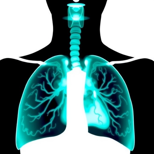
The principle behind electrical impedance tomography centers on the differential electrical conductivity of lung tissues during the breathing cycle. As air fills the alveoli, impedance changes predictably, allowing EIT to generate cross-sectional images reflecting regional ventilation. Unlike computed tomography or magnetic resonance imaging, EIT devices are compact, portable, and safe for continuous monitoring in neonatal intensive care settings. This portability facilitates its use in dynamic physiological monitoring and adjusting ventilator parameters on-the-fly, tailored individually to the infant’s lung mechanics.
Within their clinical protocol, the research team employed EIT to guide incremental lung recruitment steps during HFOV. By incrementally increasing airway pressure while observing EIT images, clinicians could optimize pressure levels to maximize alveolar recruitment while minimizing overdistension. Notably, the study highlighted significant inter-individual variability; what constitutes an optimal recruitment pressure varied considerably between infants. This finding underscores the central tenet of personalized medicine – even in the neonatal intensive care unit – to improve outcomes by customizing therapy to patient-specific physiology.
Moreover, the study’s longitudinal observations revealed that EIT-driven recruitment maneuvers correlated with improved oxygenation and more homogeneous ventilation distribution. These physiological improvements hold promise for reducing long-term pulmonary complications, such as bronchopulmonary dysplasia, which remains a major cause of morbidity in preterm survivors. As lung injury prevention becomes a cornerstone of neonatal care, real-time imaging tools like EIT are poised to become indispensable in the ventilator management arsenal.
However, while the potentials of EIT are compelling, the authors also candidly discuss limitations and practical challenges. Signal artifacts caused by electrodes or movement, as well as the current resolution constraints of EIT, require ongoing technological refinement. Furthermore, training clinicians to interpret and integrate EIT data into clinical decision-making is critical for widespread adoption. Yet, these hurdles are surmountable, and advances in artificial intelligence and machine learning could soon automate portions of image interpretation, enhancing usability and precision.
From a broader perspective, this study represents a significant leap in neonatal respiratory care by bridging technology and physiology in an elegant feedback loop. The concept of “lung-protective ventilation” is no longer theoretical but achievable in real time. By embracing continuous regional lung monitoring, the neonatal community can foresee a future where ventilator-induced injury rates decline steadily and tailored treatments become the norm rather than the exception.
The implications extend beyond the neonatal population. Insights gained from EIT monitoring during HFOV can inform adult critical care, where compromised lung mechanics demand nuanced ventilation strategies. The translational potential underscores the relevance of the research, positioning EIT at the forefront of precision ventilation monitoring across age groups and clinical contexts.
As the study illuminates, early intervention guided by accurate physiological insights delivers a dual benefit: preserving lung health while supporting survival. It captures a paradigmatic shift from reactive to proactive respiratory management. In this evolving landscape, technological innovations such as electrical impedance tomography will not only refine clinical protocols but also inspire novel therapeutic paradigms within neonatology and beyond.
Looking ahead, the researchers advocate for larger, multicenter trials to validate and expand on their promising findings. Such studies could solidify EIT’s place in evidence-based neonatal guidelines and foster widespread integration into clinical practice. Concurrent development of user-friendly interfaces and robust analytics will catalyze this transition, ultimately enhancing care quality and life quality for countless vulnerable infants worldwide.
In conclusion, the pioneering work by Werther et al. exemplifies how advanced imaging techniques can revolutionize neonatal ventilation management. By harnessing the power of electrical impedance tomography during high-frequency oscillatory ventilation, clinicians can achieve unprecedented precision in lung recruitment, safeguarding the futures of preterm infants. This research heralds a new era where bedside imaging translates directly to improved respiratory outcomes, bridging the divide between innovative technology and compassionate care.
Subject of Research: Respiratory management in preterm infants using high-frequency oscillatory ventilation guided by electrical impedance tomography.
Article Title: Preterm infants on high-frequency oscillatory ventilation: electrical impedance tomography during lung recruitment.
Article References:
Werther, T., Küng, E., Aichhorn, L. et al. Preterm infants on high-frequency oscillatory ventilation: electrical impedance tomography during lung recruitment. Pediatr Res (2025). https://doi.org/10.1038/s41390-025-04173-z
Image Credits: AI Generated
DOI: https://doi.org/10.1038/s41390-025-04173-z
Tags: dynamic lung mapping technologyEIT lung recruitment preterm infantselectrical impedance tomography benefitshigh-frequency oscillatory ventilationimproving survival rates in preterm infantsneonatal care innovationsnon-invasive imaging techniques for lungspediatric respiratory care advancementsreal-time lung ventilation visualizationreducing radiation exposure in neonatal imagingrespiratory distress syndrome in neonatesrisks of traditional ventilation strategies
