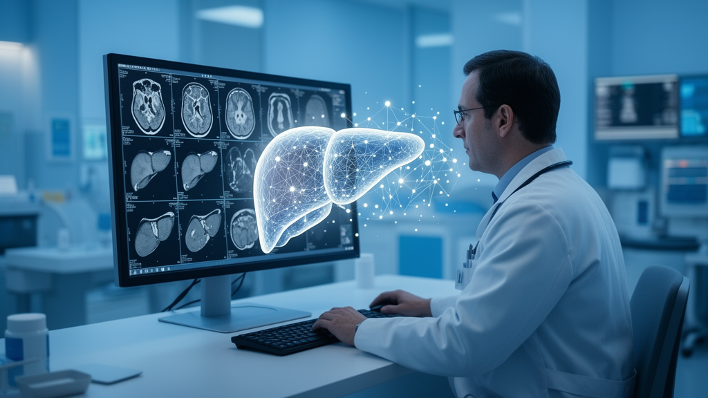
Deep learning models applied to liver MRI can detect hepatocellular carcinoma with encouraging accuracy, but the entire patient-level evidence base supporting that claim rests on just four retrospective cohorts, all conducted at Chinese tertiary centres. That is the central conclusion of a new systematic review and hierarchical meta-analysis published in the Egyptian Liver Journal, which set out to determine how mature, transportable, and clinically trustworthy the diagnostic evidence for these artificial intelligence systems actually is. The answer, according to the authors, is that the field is far less mature than the sheer volume of publications suggests, and that an evidence-maturity gap, not a new accuracy benchmark, is the study’s principal finding.
Hepatocellular carcinoma remains a major cause of cancer mortality worldwide, and clinical guidelines support structured imaging pathways for adults at risk, typically those with cirrhosis or chronic hepatitis B or C infection. Ultrasound surveillance, the mainstay of screening in these populations, has limited sensitivity for early-stage disease. Liver MRI, which combines dynamic contrast-enhanced sequences, diffusion-weighted imaging, and, when the contrast agent gadoxetate is used, hepatobiliary-phase information, generally detects hepatocellular carcinoma more sensitively than computed tomography, particularly for small lesions. Abbreviated MRI protocols may reduce the burden of examination, but subtle lesions smaller than two centimetres and variability between human readers remain important challenges. These gaps have motivated a growing literature on deep learning systems that can analyse liver MRI automatically.
The review team, led by Biruk T. Mengistie and Chernet T. Mengistie, searched MEDLINE, Embase, and Web of Science from January 2010 through July 2025, and then updated the same search strategies through 20 July 2026. The update returned 107 additional records but identified no further eligible study. From 2,235 database records in total, after deduplication and two-stage screening by independent reviewers, 28 reports were assessed in full text and 24 were excluded. Ten provided only lesion-level outcomes, six lacked reported or reconstructable patient-level data, three used a two-gate case-control design, three were non-deep-learning reports without a usable operating point, and two relied on inadequate or non-independent reference standards. Only four studies met the strict eligibility criteria, which required patient-level two-by-two accuracy data, an independent reference standard, and a defined operating point.
The four included cohorts, published between 2020 and 2025, enrolled a combined 698 adults and contributed four distinct deep learning arms: three stand-alone systems that generated outputs without radiologist input, and one radiologist-interpreted DL-assist system that altered the information available to a human reader. The review deliberately kept these paradigms separate, noting that only comparative reader studies can establish whether deep learning actually improves radiologist performance. Three of the studies used internal or same-institution testing: Lei evaluated an internal held-out test set, while Zhen and Zheng used later independent or temporal cohorts from the same institutions, which does not demonstrate geographic external validation. Only Zhang used genuine multi-centre external testing at institutions not involved in model development.
Across the four heterogeneous arms, the model-derived exploratory pooled sensitivity was 0.862 (95% confidence interval 0.819 to 0.896) and pooled specificity was 0.916 (0.881 to 0.941). Study-level sensitivity ranged from 85.1% to 93.8% and specificity from 70.0% to 100.0%. The median positive likelihood ratio, estimated by Markov chain Monte Carlo with one million draws, was 10.19 (95% interval 7.22 to 14.55), the negative likelihood ratio was 0.151 (0.114 to 0.199), and the diagnostic odds ratio was 67.59 (41.10 to 111.10). The authors stress repeatedly that these are exploratory model summaries, not stable population-performance benchmarks, because four studies cannot support reliable variance, subgroup, or meta-regression inference.
A striking statistical feature of the synthesis underscores that caution. The estimated between-study variances for logit sensitivity and logit false-positive rate lay at the boundary of the parameter space, at 1.60 times ten to the minus nine and 2.56 times ten to the minus eight respectively, producing confidence and prediction regions that were nearly superimposed. The authors explicitly warn that these boundary values and numerically small I-squared analogues are uninformative with only four studies and must not be interpreted as evidence of statistical or clinical homogeneity. The included studies differed materially in clinical role, testing provenance, MRI acquisition and preprocessing, lesion spectrum, and reference standards, differences that no subgroup analysis could resolve because none was attempted.
The methodological quality assessment was equally sobering. Using the QUADAS-2 tool, two independent reviewers judged patient selection to be at high risk of bias in two of four studies and unclear in the other two; the index test domain was low risk in only one study; the reference standard was unclear in all four; and flow and timing was high risk in three of four. These judgments reflect selected and sometimes small-lesion-enriched spectra, image-quality exclusions, incompletely locked or blinded index tests, and differential or composite verification in which HCC cases were often confirmed by histology while non-HCC participants were classified by imaging or follow-up, raising the possibility of incorporation bias when index-MRI features contributed to the imaging-based reference classification.
Reporting quality for artificial intelligence-specific items was also incomplete. Although all four studies reported dataset sources and train-test splits, external testing under a definition requiring evaluation at a different institution was reported in only one study, explainability in one, code or model availability in one, and workflow integration in one. Regulatory status was reported in none. After reclassifying the two same-centre temporal cohorts, overall AI-reporting ratings were Good for one study, Moderate for one, and Poor for two. Technical details such as coil configuration, reconstruction software, missing-sequence handling, and model failure reporting were usually incomplete, and the studies also diverged in contrast protocols: only one used acquired gadoxetate hepatobiliary-phase images, one synthesized contrast phases from precontrast data as model outputs rather than acquired images, and one did not explicitly report hepatobiliary-phase acquisition.
The clinical implications follow directly from these limitations. Applying the pooled median likelihood ratios to selected pretest probabilities produces illustrative post-test probability shifts, but the authors emphasize that these are mathematical examples, not externally validated decision thresholds, and that they do not demonstrate clinical utility. They caution that a negative model output must not independently terminate assessment in a high-risk patient. Because all four studies originated from Chinese tertiary centres, often in hepatitis B-predominant high-risk populations, transportability across aetiologies, demographics, community settings, vendors, field strengths, acquisition protocols, and image-quality conditions is unknown. No included study performed a cost-effectiveness or budget-impact evaluation, and none established whether deep learning augments radiologist performance.
The review’s conclusion is measured rather than dismissive: deep learning applied to liver MRI produced promising model-derived patient-level summaries, but four retrospective, geographically concentrated cohorts do not establish a transportable accuracy benchmark, radiologist augmentation, or clinical benefit. The authors call for prospective multi-centre, multi-vendor testing, paired or randomized reader studies comparing radiologist-alone against DL-assist reading, locked thresholds and calibration, explicit blinding and failure reporting, domain-shift testing across hardware and protocols, live workflow monitoring, and proportionate economic evaluation. Until such studies exist, they argue, the field’s most important product is not an accuracy figure but a clearly documented gap between publication volume and clinically usable evidence.
Subject of Research: Diagnostic accuracy of deep learning applied to liver MRI for detecting hepatocellular carcinoma
Article Title: Diagnostic accuracy of deep learning applied to liver MRI for hepatocellular carcinoma detection: a systematic review and HSROC meta-analysis of patient-level evidence
Article References: Mengistie, B. T., Gete, K. Y., Mohammed, A., Zimme, Z. A., Teferi, M. G., Adugna, A., Kibret, F., Fetle, Y., Abera, B. F., & Mengistie, C. T. (2026). Diagnostic accuracy of deep learning applied to liver MRI for hepatocellular carcinoma detection: a systematic review and HSROC meta-analysis of patient-level evidence. Egyptian Liver Journal, 16(1), Article 74. https://doi.org/10.1186/s43066-026-00546-z
Image Credits: AI Generated
DOI: 10.1186/s43066-026-00546-z
Keywords: deep learning, liver MRI, hepatocellular carcinoma, diagnostic accuracy, systematic review, meta-analysis, HSROC, QUADAS-2, external validation, radiology AI, evidence quality, medical imaging
News Source: Ophelia Keating. (October 28, 2026). Deep Learning on Liver MRI Shows Promise for Detecting Liver Cancer, but Evidence Remains Thin. Scienmag.