


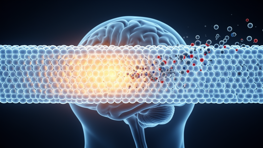
For decades, drug developers have treated the blood-brain barrier as the great wall of the central nervous system: once a therapeutic molecule got past it, the battle was assumed to be won. A new Mini Review published in Frontiers in Drug Discovery argues that this assumption is fundamentally flawed. Written by Nagisa Arimitsu Nakata, Masaaki Kurita, and colleagues at St. Marianna University School of Medicine in Japan, the review reframes CNS drug delivery as a spatial and dynamic pharmacokinetic process rather than a single-step event of barrier penetration. Even after a drug crosses brain capillary endothelial cells, the authors contend, it must still navigate perivascular compartments, extracellular diffusion barriers, non-target cellular uptake, and intracellular trafficking before it can reach its molecular target in an active form.
The distinction matters because successful BBB penetration does not necessarily ensure effective therapeutic action within the brain. The authors point to Alzheimer’s disease, where anti-amyloid beta antibodies may show evidence of brain entry or target engagement, yet this does not always translate into sufficient clinical efficacy. Such outcomes suggest that CNS delivery should not be defined solely by BBB permeability or by total brain concentration. Instead, after crossing the barrier, drugs must traverse a series of cellular and intracellular barriers before reaching the intended molecular target, and a substantial gap can open between apparent brain exposure and effective target-site exposure.
The review lays out these post-BBB obstacles as a sequence of spatial filtering steps. One key compartment is the perivascular space, where molecules that cross or bypass the BBB may linger rather than immediately entering the parenchyma. This restriction is not explained by a single size-exclusion mechanism; parenchymal access reflects a balance among molecular size, diffusivity, interactions with perivascular and extracellular structures, and redistribution through paravascular fluid pathways. Large macromolecules, including antibodies, diffuse slowly through brain interstitial spaces and may remain concentrated near vascular or perivascular compartments before achieving deeper penetration.
Recent studies using whole-brain tissue clearing and cell-type-resolved analyses sharpen this picture. Control IgG was found to localize predominantly to perivascular and leptomeningeal compartments, with minimal distribution to parenchymal cells, whereas antibody transport vehicles targeting the transferrin receptor or the CD98 heavy chain achieved substantially broader parenchymal and cell-type distribution even when bulk brain exposures were comparable. In another recent study, fusing a transferrin receptor 1-binding BBB shuttle to aducanumab produced enhanced and more homogeneous whole-brain distribution and reduced periarterial accumulation compared with unmodified aducanumab. These findings provide direct evidence that spatial distribution within the CNS can differ markedly despite similar or apparently successful brain exposure.
Beyond the perivascular interface, astrocytic endfeet, the glia limitans, basement membrane components, and the extracellular matrix create a highly organized microenvironment that restricts movement into the parenchyma. The tortuous brain extracellular space then imposes size- and structure-dependent constraints on intraparenchymal diffusion, particularly for macromolecules and nanoscale therapeutics. For nanoparticulate systems, tissue penetration depends not only on particle size but also on surface properties such as charge, coating density, and ligand presentation; dense polyethylene glycol coatings, for example, can reduce adhesive interactions with extracellular components and improve nanoparticle penetration through brain tissue.
At the cellular level, a further filter awaits. Once a therapeutic agent reaches the parenchyma, it may be preferentially internalized by non-target cells, including microglia, astrocytes, or endothelial-associated cells, thereby reducing the fraction available to the intended target cell. The review describes this “sink effect” as particularly relevant to macromolecular and nanoparticulate systems that undergo receptor-mediated or phagocytic uptake. Under such conditions, whole-brain homogenate analysis may indicate high brain exposure even though the actual amount of active drug reaching target neurons, tumor cells, or disease-relevant immune cells remains insufficient.
Intracellular barriers are not confined to large molecules. Certain weakly basic and lipophilic small molecules accumulate in acidic lysosomal compartments through ion trapping. Sunitinib and palbociclib are cited as well-characterized examples in which lysosomal sequestration increases total intracellular drug accumulation while reducing the freely available cytosolic fraction capable of engaging intracellular targets. The authors caution that this phenomenon is compound dependent and may be partially overcome at higher extracellular concentrations, so it should not be regarded as a universal limitation of small-molecule CNS delivery. Rather, it illustrates how total cellular drug accumulation can become uncoupled from pharmacologically active intracellular exposure. For macromolecular therapeutics, intracellular trafficking similarly determines whether internalized molecules undergo recycling, productive transport, or lysosomal degradation.
These barriers also undermine a familiar pharmacological assumption: that raising the dose raises the effect. In CNS drug delivery, the relationship is frequently nonlinear. A higher plasma concentration does not necessarily produce proportional increases in effective exposure within the parenchyma or at the intended molecular target, because delivery is shaped by saturable transport systems, endothelial retention, cell-type-specific uptake, and intracellular sequestration. The maximum tolerated dose may therefore not correspond to the optimal biological dose for CNS therapeutics.
Receptor-mediated transcytosis offers a concrete illustration. For antibody-based delivery systems targeting the transferrin receptor, brain uptake is not simply improved by stronger receptor binding or higher systemic exposure. High-affinity or excessive receptor engagement can promote retention within brain endothelial cells and reduce efficient release to the parenchymal side, limiting functional delivery despite apparent BBB targeting. In this context, the review notes, the BBB endothelium may act not only as a transport interface but also as a site of sequestration or degradation, particularly when receptor-mediated pathways become saturated or biased toward intracellular retention.
Given these limitations of bulk measurements, the review devotes substantial attention to imaging-based evaluation of spatial CNS pharmacokinetics. Conventional approaches relying on plasma concentration, CSF concentration, or total brain homogenate analysis are useful for estimating overall exposure but cannot determine where a drug is located within the CNS. Each modality carries trade-offs. PET and SPECT are clinically translatable and suitable for whole-brain pharmacokinetic evaluation, but their spatial resolution is insufficient to resolve cellular-level delivery barriers. Two-photon intravital microscopy enables real-time visualization of vascular and cellular dynamics at high spatial resolution, but its imaging depth is limited and it often requires fluorescent labeling. MALDI mass spectrometry imaging can map molecules according to mass, distinguishing parent compounds from metabolites in tissue sections, but it requires tissue sections and provides no real-time information. No single modality is sufficient; the authors argue for multimodal strategies matched to the stage of drug development.
Quantitative radiotracing studies underscore why molecular identity matters. In a recent study comparing residualizing zirconium-89 and non-residualizing iodine-125 labels on TfR1- and CD98hc-targeted bispecific antibodies, apparent brain-associated radioactivity was strongly influenced by cellular internalization and catabolism. For a TfR1-targeted bispecific antibody engaging the internalizing neuronal receptor TrkB, brain signals measured with the two isotopes diverged by more than 30-fold. The authors highlight this as evidence that intact, pharmacologically relevant therapeutic molecules must be distinguished from signals generated by intracellular uptake and degradation products when evaluating CNS exposure. They also flag a technical caveat: fluorescent dyes or bulky probes can alter the physicochemical properties of small molecules, so labeled compounds may not faithfully represent the distribution of the original drug. Label-free approaches such as stimulated Raman scattering microscopy and mass spectrometry imaging may help address this.
The review then turns to temporal modifiers of CNS barriers. Barrier function is dynamically regulated over time, not a fixed anatomical property. The glymphatic system, which contributes to the exchange and clearance of solutes within the brain, shows state-dependent and circadian variation; glymphatic and lymphatic fluid flow has been reported to fluctuate according to sleep-wake state and circadian timing. This raises the possibility that the timing of drug administration could influence how long drugs remain within the brain interstitium. For drugs requiring sustained parenchymal retention, enhanced clearance may reduce effective exposure; conversely, when clearance itself is therapeutically desirable, such as in strategies aimed at promoting removal of pathological proteins, synchronization with glymphatic activity may be beneficial. Efflux transporters such as P-glycoprotein are also subject to circadian regulation, suggesting that specific dosing windows may exist in which efflux activity, parenchymal entry, or drug retention differs across the day, although the authors note this concept requires further validation for individual drugs and disease settings.
Glymphatic function is also disease dependent and pharmacologically modifiable. In an experimental model of intraventricular hemorrhage, disruption of glia-vascular unit integrity through c-Src/beta-dystroglycan signaling was associated with loss of perivascular aquaporin-4 polarization, impaired glymphatic function, and secondary hydrocephalus. Conversely, therapeutic hypothermia has recently been reported to enhance drainage through the glymphatic-meningeal lymphatic-deep cervical lymphatic pathway and to improve hydrocephalus and neurological dysfunction after experimental intraventricular hemorrhage. Although these studies do not directly examine CNS drug delivery, they show that glymphatic transport can change with disease and respond to intervention.
Aging represents a longer-term temporal modifier. Normal aging is associated with BBB dysfunction, particularly in vulnerable regions such as the hippocampus, and with impairment of paravascular or glymphatic clearance pathways. These changes may have complex and sometimes opposing effects: a more permeable BBB may increase apparent brain entry, while impaired clearance may prolong intraparenchymal retention and increase the risk of local toxicity. Studies using only young, healthy animal models may therefore fail to capture delivery kinetics relevant to elderly patients, who represent a major population for many CNS disorders.
Translational considerations compound the challenge. Many high-resolution imaging approaches are performed primarily in rodents, yet substantial anatomical and physiological differences exist between rodent and human brains, including brain volume, white-to-gray matter composition, vascular architecture, interstitial transport distances, and fluid-clearance dynamics. Classic distribution data illustrate the point: autoradiograms of rat brain prepared 20 hours after a single intracerebroventricular injection of radiolabeled brain-derived neurotrophic factor showed distribution exclusively along the ependymal surface of the injected ventricle, and in primates, concentrations of water-soluble small molecules fell approximately 10-fold per millimeter of distance from the CSF surface, a decline even steeper for macromolecules. Human MRI studies using intrathecal tracers have begun to define these dynamics clinically, with first detectable tracer enrichment at approximately 1 hour in cortical regions but after approximately 2 to 4 hours in corresponding white matter. Disease adds further heterogeneity: in gliomas, vascular disruption is spatially heterogeneous, and even after extravasation, abnormal tumor architecture and elevated interstitial fluid pressure can restrict diffusion, so increased vascular leakiness does not ensure therapeutically sufficient concentrations throughout the tumor.
Subject of Research: Post-BBB spatial and dynamic barriers that govern CNS drug delivery and target engagement
Article Title: Beyond BBB permeability: spatial and dynamic post-BBB barriers governing CNS drug action
Article References: Nakata, N. A., Kurita, M., Ohashi, A., Matsumoto, K., Miyabe, C., Murata, H., & Miyabe, Y. (2026). Beyond BBB permeability: spatial and dynamic post-BBB barriers governing CNS drug action. Frontiers in Drug Discovery, 6, Article 1957594. https://doi.org/10.3389/fddsv.2026.1957594
Image Credits: AI Generated
DOI: 10.3389/fddsv.2026.1957594
Keywords: blood-brain barrier, CNS drug delivery, perivascular space, glymphatic system, mass spectrometry imaging, intravital microscopy, lysosomal sequestration, receptor-mediated transcytosis, nanoparticles, Alzheimer's disease, pharmacokinetics, aging
News Source: Cassandra Pierce. (October 29, 2026). Crossing the Blood-Brain Barrier Is Only the First Step for CNS Drugs. Scienmag.



