Down syndrome, caused by an extra copy of chromosome 21, has long been studied through the lens of gene dosage, yet the cellular machinery that actually fails in trisomy 21 has remained stubbornly opaque. A new study published in Frontiers in Genetics has now traced a surprising thread connecting the centromere—the chromosomal structure that governs cell division—to the energy factories of the cell. Researchers at Shanghai Children’s Hospital and Shanghai Jiao Tong University School of Medicine, working with colleagues at Fudan University, report that an immune protein called IFIT3, when overproduced, is sufficient to impair mitochondrial function and disrupt cell cycle progression in human stem cells, recapitulating hallmark cellular defects seen in Down syndrome.
The research team, led by Jing-bin Yan and Fanyi Zeng, focused on a question that has received surprisingly little attention: what happens to the proteins that cluster around centromeres in fetal cells carrying three copies of chromosome 21? Centromeres are not merely passive anchors for spindle fibers during cell division. They coordinate kinetochore assembly, microtubule attachment, and checkpoint signaling, and their dysfunction is known to destabilize the genome. Because chromosome 21 non-disjunction—the meiotic error that produces trisomy 21—depends entirely on intact centromere-kinetochore function, the investigators reasoned that the centromere-proximal proteome might hold clues to how the extra chromosome wreaks havoc on cellular physiology.
To map this proteomic landscape, the team employed a sophisticated technique called APEX2 proximity labeling. They engineered a fusion protein combining CENPA, the defining histone variant of centromeres, with GFP and the engineered peroxidase APEX2. When expressed in living cells and activated with hydrogen peroxide, APEX2 covalently tags every protein within roughly 20 nanometers of the centromere with biotin. These tagged proteins can then be pulled down with streptavidin beads and identified by quantitative mass spectrometry. Unlike conventional co-immunoprecipitation, which can lose weak or transient interactions and disrupt subcellular compartments, proximity labeling preserves the native molecular environment of the centromere in living cells.
The researchers applied this system to primary human amniocytes—cells shed by the fetus into amniotic fluid—obtained from pregnant women undergoing prenatal diagnosis at the Shanghai Ji Ai Genetics and IVF Institute. They compared trisomy 21-derived amniocytes with euploid normal amniocytes matched for gestational age, fetal sex, and passage number. Karyotyping confirmed the chromosomal constitution of each sample, and all experiments used cells at passage five or earlier to avoid senescence-related artifacts. After validating that the CENPA-GFP-APEX2 fusion protein localized exclusively to centromeres and exhibited minimal background biotinylation, the team proceeded to quantitative proteomic profiling.
Using label-free liquid chromatography-tandem mass spectrometry, the investigators quantified 999 CENPA-proximal proteins across three biological replicates per group. Among these, 32 proteins showed significantly altered abundance in trisomy 21 amniocytes: 19 were upregulated and 13 were downregulated. Principal component analysis revealed tight clustering within each group and clear separation between the two conditions, indicating widespread proteome remodeling driven by the extra chromosome. Gene Ontology and Gene Set Enrichment analyses pointed to dysregulation of interferon response pathways and ribosomal protein networks, while Kyoto Encyclopedia of Genes and Genomes pathway analysis implicated thyroid hormone signaling, platelet activation, and RAP1 signaling among the perturbed pathways.
Standing out among all the differentially abundant proteins was IFIT3, or Interferon-induced protein with tetratricopeptide repeats 3, which showed the most significant upregulation in trisomy 21 amniocytes. The team validated this finding by Western blot using independent donor-matched samples, confirming that IFIT3 protein levels were indeed elevated in the trisomy 21 cells. IFIT3 is a canonical interferon-stimulated gene, part of a family of antiviral effector proteins that are chronically hyperactivated across all trisomy 21 cell types—a phenomenon sometimes referred to as the interferonopathy of Down syndrome. What made the detection of IFIT3 within the centromere-proximal proteome particularly intriguing is that this protein is predominantly cytoplasmic at steady state, suggesting it may be transiently recruited to pericentromeric chromatin during mitosis.
To determine whether elevated IFIT3 is merely a bystander or an active driver of cellular dysfunction, the researchers generated human induced pluripotent stem cells overexpressing IFIT3 via lentiviral transduction. Before conducting any functional assays, they confirmed that the engineered cells retained intact pluripotency, with normal expression of OCT4, NANOG, SOX2, and SSEA4. The results were striking. Forced IFIT3 overexpression triggered G1-phase cell cycle arrest, suppressed cell proliferation, and elevated apoptotic rates in the pluripotent stem cells. Western blot analysis revealed that P21 and P27, two master negative regulators of cell cycle progression, were substantially downregulated in primary trisomy 21 amniocytes, providing a molecular link between IFIT3 dysregulation and cell cycle perturbation.
The most consequential findings, however, concerned mitochondrial function. Transcriptome-wide RNA sequencing of IFIT3-overexpressing stem cells identified 200 differentially expressed genes, with strong transcriptional repression of oxidative phosphorylation and mitochondrial ATP biogenesis gene sets. Gene Set Enrichment Analysis independently validated suppressed mitochondrial respiration signatures alongside enrichment of axonal guidance and neurogenesis gene sets. The team then performed direct functional measurements using JC-1 staining to assess mitochondrial membrane potential and luminescence-based assays to quantify intracellular ATP. Compared with control cells, IFIT3-overexpressing stem cells displayed a significantly reduced red-to-green fluorescence ratio, indicating depolarized mitochondria, and approximately 20 percent lower intracellular ATP levels. These results confirmed that IFIT3 overexpression alone is sufficient to impair mitochondrial bioenergetics.
The study’s authors note an intriguing discrepancy: forced IFIT3 overexpression in pluripotent stem cells induced G1-phase arrest, whereas endogenous IFIT3 upregulation in primary trisomy 21 amniocytes promoted increased S-phase entry. They attribute this to cell-type-specific divergence in cell cycle checkpoint wiring between differentiated primary amniocytes and pluripotent stem cells, highlighting the context-dependent nature of IFIT3’s mitotic regulatory functions. This nuance underscores the complexity of translating findings across cellular models and suggests that IFIT3’s effects on cell cycle progression may depend on the developmental stage and differentiation status of the cell.
Looking forward, the researchers propose that pharmacological suppression of IFIT3 activity may restore mitochondrial bioenergetics and reverse core cellular pathologies in Down syndrome. This builds on prior preclinical evidence that 7,8-dihydroxyflavone, a small molecule that ameliorates mitochondrial dysfunction, can rescue neurodevelopmental defects in the Ts65Dn mouse model of trisomy 21. The current work distinguishes itself from a previous study by the same group, which showed that the chromosome 21-encoded transcription factor RUNX1 disrupts mitochondrial function through PI3K-Akt signaling. The present findings identify a genetically independent, interferon-dependent IFIT3 signaling axis that acts upstream of mitochondrial impairment, providing a novel proteomic framework that links centromere-proximal immune signaling to bioenergetic failure in trisomy 21 and nominating IFIT3 as a candidate therapeutic target for future intervention studies.
Subject of Research: The role of centromere-proximal IFIT3 overexpression in mitochondrial dysfunction and cell cycle disruption in Down syndrome
Article Title: Overexpression of centromere-localized IFIT3 impairs mitochondrial function and disrupts cellular proliferation and cell cycle progression in down syndrome
Article References: Overexpression of centromere-localized IFIT3 impairs mitochondrial function and disrupts cellular proliferation and cell cycle progression in down syndrome. (n.d.). https://doi.org/10.3389/fgene.2026.1904151
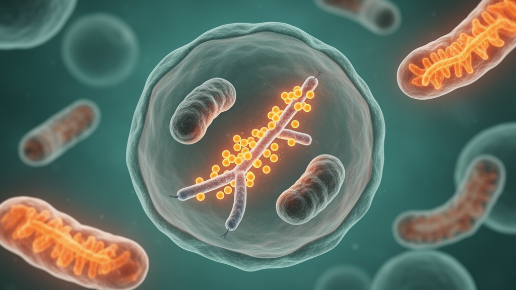
Image Credits: AI Generated
DOI: 10.3389/fgene.2026.1904151
Keywords: Down syndrome, trisomy 21, IFIT3, centromere, CENPA, mitochondrial dysfunction, APEX2 proximity labeling, proteomics, oxidative phosphorylation, cell cycle, induced pluripotent stem cells, interferon signaling
News Source: Drew Townsend. (October 16, 2026). Centromere Protein IFIT3 Emerges as a Driver of Mitochondrial Dysfunction in Down Syndrome. Scienmag.