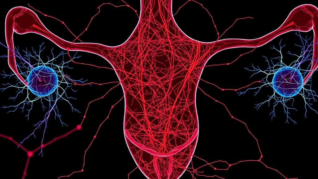
Endometriosis has long been one of medicine’s most frustrating puzzles: a disease that affects roughly one in ten individuals assigned female at birth, causes chronic pain that can be debilitating, and yet whose underlying biology remains stubbornly poorly understood. Now a team at The Jackson Laboratory for Genomic Medicine and UConn Health has produced the most detailed spatial atlas to date of human endometriosis lesions, mapping, cell by cell, the molecular conversations taking place inside ovarian and peritoneal lesions. The study, published in Nature Communications, reveals shared architectural features across lesion types and points to specific neuroimmune interactions that may help explain why the disease hurts so much.
The research, led by Caroline M. Haney and Elise T. Courtois, with Danielle E. Luciano and colleagues, tackles a problem that has hampered endometriosis research for decades. Endometriosis occurs when tissue resembling the uterine lining grows outside the uterus, most commonly on the ovaries and the peritoneum, the membrane lining the abdominal cavity. These lesions behave like small, inflamed organs in their own right, complete with glandular epithelium, supporting stromal cells, immune infiltrates and, as this study emphasizes, nerve fibers. Traditional bulk sequencing methods average out the signals from all of these cell types, obscuring the local interactions that likely drive symptoms.
Spatial transcriptomics changes that calculus fundamentally. Rather than dissociating tissue into a soup of single cells, the technology measures gene expression directly in intact tissue sections, preserving the geographic relationships between cells. In this study, the team applied high-resolution spatial profiling to human ovarian and peritoneal endometriosis specimens, effectively producing a molecular cartography of each lesion. Every cell’s identity and gene expression profile could be located within the tissue landscape, allowing the researchers to ask not just which cells are present, but which cells sit next to which, and what signaling molecules they exchange at those interfaces.
The resulting maps revealed striking commonalities between lesion types that are often treated as clinically distinct. Both ovarian and peritoneal lesions showed immune cell infiltration, organized fibroblast compartments encircling the epithelial glands, and characteristic distributions of neuronal and macrophage subsets. This shared spatial blueprint suggests that despite their different locations, the two most common lesion types may operate through convergent biological mechanisms, a finding with real implications for how therapies are designed and tested. A drug targeting a pathway common to both lesion environments could plausibly benefit patients regardless of where their disease manifests.
One of the most consequential contributions of the study is its precise definition of the sensory neuronal subtypes present within lesions and the mapping of their positions relative to immune cells. Pain in endometriosis is the dominant clinical complaint, yet the neural wiring of lesions has been difficult to study in human tissue. By identifying which classes of sensory neurons innervate the lesions and demonstrating that they occupy defined spatial relationships with macrophages and other immune populations, the researchers have built a structural case for neuroimmune crosstalk as a driver of endometriosis-associated pain. Macrophages, the tissue-resident immune cells capable of releasing inflammatory mediators and growth factors, are known to modulate nerve sensitivity in other chronic pain conditions, and the spatial proximity documented here suggests a similar dynamic may be at work in endometriotic tissue.
The fibroblast findings may prove equally important. Fibroblasts, the connective tissue cells that form the stromal scaffold of organs, were found to form distinct compartments surrounding the epithelial glands within lesions. In many fibrotic and inflammatory diseases, activated fibroblasts secrete cytokines, extracellular matrix components and growth factors that reshape the local environment and sustain chronic inflammation. The observation that these cells organize into periglandular niches in endometriosis lesions, conserved across lesion types, suggests they may be active participants in lesion maintenance rather than passive structural support, potentially creating the microenvironment in which epithelial cells, immune cells and nerves interact.
Crucially, the team did not stop at descriptive mapping. To test whether the epithelial-neuronal interactions inferred from the tissue sections are real, functional relationships, they built an in vitro model combining 3D peripheral sensory brain organoids with human endometriosis epithelial and fibroblast cells. Organoids are lab-grown, three-dimensional tissue cultures that recapitulate key features of the organ or tissue they model; sensory organoids derived from human induced pluripotent stem cells can generate neurons with the properties of peripheral sensory nerves. By co-culturing these neural structures with endometriosis-derived epithelial and stromal cells, the researchers could validate the interactome, the network of signaling interactions, that their spatial maps had predicted, providing experimental support that the epithelial-neuronal communication observed in patient tissue is biologically meaningful.
The technical achievement underlying these results should not be understated. The study drew on the EndoRISE Biorepository, part of the Connecticut Endometriosis Tissue and Data Biorepository funded by the State of Connecticut and The Mayday Fund, which supplied the patient biospecimens essential for such high-resolution work. The analysis leveraged The Jackson Laboratory’s Single Cell Biology Lab, Genome Technologies, Histology and Microscopy cores, along with high-performance computing resources, and used induced pluripotent stem cell lines from the laboratory’s Genetic Discovery initiative for the organoid work. Studies of this scale depend on patients consenting to donate tissue during surgery, and the authors specifically credit the study participants and the surgical teams at UConn Health who made the specimen collection possible.
What makes the study resonate beyond the technical community is what it implies for patients. Current endometriosis treatment options, hormonal suppression and surgical excision, address symptoms or remove visible disease but do not target the specific cellular mechanisms that generate pain and drive progression, and recurrence after surgery is common. A spatially resolved, mechanistic understanding of the lesion microenvironment gives drug developers concrete targets: the signaling pathways between sensory neurons and macrophages, the fibroblast niches around epithelial glands, and the conserved features shared across lesion types. Neuroimmune interactions in particular have emerged as a promising frontier in chronic pain research more broadly, and this study provides disease-specific, human-tissue evidence for pursuing that frontier in endometriosis.
There are, of course, limits to what any single atlas can establish. The study maps two lesion types, ovarian and peritoneal, and the authors frame their work as establishing the spatial transcriptomic cartography of those lesions rather than a complete account of all endometriosis presentations, such as deeply infiltrating disease. The organoid co-culture system validates epithelial-neuronal interactions in vitro but remains a simplified model of a complex in vivo environment. Still, the combination of high-resolution human tissue mapping with functional validation represents a methodological template that other chronic inflammatory diseases could follow. For a condition that has historically been underfunded, under-researched and frequently dismissed, the appearance of a rigorous, open-access, spatially resolved molecular atlas of endometriosis lesions marks a genuine step forward, one that transforms the disease from an opaque clinical syndrome into a mappable biological landscape with identifiable cellular players and testable therapeutic targets.
Subject of Research: Spatial transcriptomic mapping of fibroblast and neuroimmune microenvironments in human endometriosis lesions
Article Title: High-resolution spatial transcriptomics reveals fibroblast and neuroimmune microenvironments in endometriosis lesions
Article References: Haney, C. M., Alizadeh, E., Lee, J., Sullivan, M., Kuljancic, J., Flynn, W. F., Robson, P., White, B. S., Luciano, D. E., & Courtois, E. T. (2026). High-resolution spatial transcriptomics reveals fibroblast and neuroimmune microenvironments in endometriosis lesions. Nature Communications. https://doi.org/10.1038/s41467-026-78453-5
Image Credits: AI Generated
DOI: 10.1038/s41467-026-78453-5
Keywords: endometriosis, spatial transcriptomics, fibroblasts, sensory neurons, macrophages, neuroimmune interactions, chronic pain, organoids, ovarian lesions, peritoneal lesions, single-cell biology, Nature Communications
News Source: Ophelia Keating. (October 11, 2026). Cell Maps of Endometriosis Reveal Nerve and Immune Hubs That May Drive Pain. Scienmag.