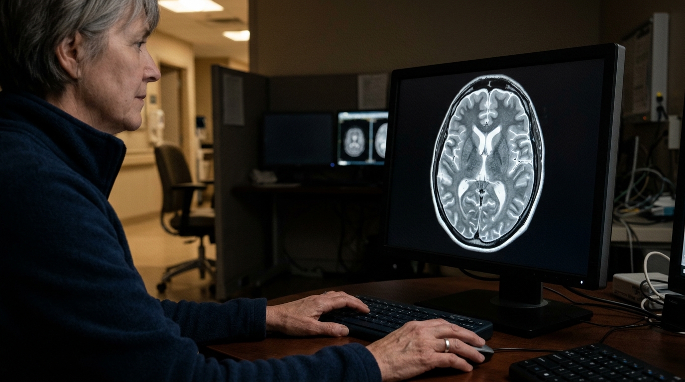
A large retrospective study of patients with extensive strokes affecting at least half of the middle cerebral artery territory has found that brains with more atrophy are substantially less likely to develop severe, life-threatening mass effect. The research, published in Neurocritical Care, analyzed 565 patients admitted to two academic hospitals with comprehensive stroke centers between 2006 and 2024, and showed that greater quantified brain atrophy was associated with lower odds of midline shift of 5 millimeters or more on follow-up head CT, with an odds ratio of 0.44 and a 95 percent confidence interval of 0.34 to 0.58. Notably, atrophy was not associated with in-hospital mortality, suggesting its prognostic value is specific to the radiographic course of cerebral edema rather than to survival overall.
Large middle cerebral artery infarctions can trigger malignant cerebral edema, a swelling of brain tissue that displaces structures across the midline and can become fatal without timely intervention. Clinicians currently rely on established clinical and radiographic predictors to gauge which patients are most at risk, but these tools leave considerable uncertainty. The research team, led by Yili Du and Charlene J. Ong along with colleagues including Aiman Z. Altaf, Nawal J. Ibrahim, Stefanos Chatzidakis, Huimin Cheng, David M. Greer, Rajat Dhar and others, hypothesized that the amount of brain tissue already lost to age-related shrinkage might meaningfully alter how much room the brain has to accommodate swelling after a major infarct. To test this, they quantified atrophy as the inverse of standardized brain volume measured on the admission head CT, turning a familiar but often subjective radiographic impression into a numerical biomarker.
The study enrolled adults who presented within 24 hours of their last known well time and who had at least one follow-up head CT during their hospital stay. The primary outcome was severe radiographic mass effect, defined as midline shift of at least 5 millimeters on follow-up imaging. The secondary outcome was in-hospital mortality. Because the design was a retrospective observational cohort, the findings describe associations rather than proven causal effects, and the study was approved by the institutional review boards of both participating centers.
Among the 565 patients, whose mean age was 67.5 years with a standard deviation of 15.7, and of whom 49.9 percent were female, 223 patients, or 39.5 percent, went on to develop severe mass effect. In multivariable regression models, greater atrophy remained independently associated with lower odds of reaching that 5-millimeter midline shift threshold. The direction of the association may seem counterintuitive at first glance, since one might assume a smaller brain would be more vulnerable to swelling. The authors interpret the result within the framework of intracranial compliance: a brain that has already lost volume may have more residual intracranial space relative to the volume of expanding edema, allowing displacement to accumulate before it crosses the severe threshold.
Beyond the association itself, the team asked whether adding atrophy to existing predictive models actually improves their performance. They compared multivariable models with and without the atrophy measure using measures of goodness of fit, calibration, and discrimination. The model including atrophy showed a substantially better fit, with a likelihood ratio test yielding a chi-squared statistic of 41 on one degree of freedom and a p value below 0.001. Information-theoretic criteria also favored the expanded model, with the Akaike information criterion improving from 741 to 703 and the Bayesian information criterion from 767 to 733. Discrimination improved as well, with the area under the receiver operating characteristic curve rising from 0.60 in the baseline model to 0.68 once atrophy was incorporated.
These gains are meaningful in a clinical landscape where early risk stratification directly shapes monitoring intensity and decisions about interventions such as decompressive hemicraniectomy. An AUC of 0.68 remains moderate in absolute terms, and the study does not claim that atrophy alone can predict outcomes; rather, the value lies in incremental improvement over established clinical and radiographic predictors already in use. The authors conclude that quantified brain atrophy is independently associated with a reduced risk of severe mass effect after large MCA stroke and that incorporating this imaging biomarker may enhance early risk stratification, monitoring, and intervention planning for patients at risk of life-threatening cerebral edema.
Several limitations temper the strength of the conclusions. The retrospective design means atrophy and outcomes were measured from existing records rather than through a prospectively defined protocol, and the cohort was drawn from two academic comprehensive stroke centers, which may limit generalizability to broader practice settings. The follow-up CT requirement introduces the possibility of selection effects, since patients who deteriorate rapidly or die before repeat imaging may be underrepresented. The mortality finding, or lack thereof, is particularly worth emphasizing: while atrophy predicted radiographic mass effect, it did not predict in-hospital death, so clinicians should not read the biomarker as a survival prognosticator. The odds ratio describes association within this cohort and does not establish that preserving brain volume would prevent mass effect.
What the study does offer is a concrete, measurable variable that refines an existing clinical dilemma. Malignant edema after large hemispheric infarction follows a trajectory that clinicians must anticipate within hours to days, and tools that sharpen early stratification can inform decisions about when to escalate monitoring or prepare for surgical intervention. The finding that a routinely obtainable CT-derived measure adds statistically significant predictive value suggests a practical avenue for integrating brain volume quantification into standard risk models, though prospective validation in more diverse cohorts would be the natural next step before widespread clinical adoption.
Subject of Research: The association between quantified brain atrophy and severe mass effect after large middle cerebral artery ischemic stroke.
Article Title: Quantified Brain Atrophy and Risk of Severe Mass Effect in Acute Ischemic Stroke
Article References: Du, Y., Altaf, A. Z., Ibrahim, N. J., Chatzidakis, S., Mallinger, L. A., Reinert, A. L., Stafford, R. A., Kumar, A., Avula, A., Abdalkader, M., Cheng, H., Greer, D. M., Dhar, R., & Ong, C. J. (2026). Quantified Brain Atrophy and Risk of Severe Mass Effect in Acute Ischemic Stroke. Neurocritical Care. https://doi.org/10.1007/s12028-026-02660-7
Image Credits: AI Generated
DOI: 10.1007/s12028-026-02660-7
Keywords: brain atrophy, acute ischemic stroke, middle cerebral artery, cerebral edema, midline shift, mass effect, head CT, risk stratification, Neurocritical Care, predictive modeling, intracranial compliance, observational cohort
News Source: Cassandra Pierce. (November 5, 2026). Brain Atrophy Lowers Odds of Severe Midline Shift After Large Stroke. Scienmag.