Cartilage loss and thinning bones are often studied side by side in the same joint, but an analysis of the human femoral head shows that the two disease processes leave their fingerprints at different depths of the bone. In a study published in Arthritis Research & Therapy, researchers examined 30 femoral heads removed during primary total hip arthroplasty for hip osteoarthritis and mapped bone microarchitecture layer by layer, from the subchondral mineralized plate at the articular surface down to the deepest trabecular core. Their central finding is a spatial division of labor: cartilage degeneration independently predicted subchondral bone structure only in the plate and in superficial to intermediate trabecular regions extending to 13.12 millimeters in depth, while systemic bone mineral density emerged as an independent predictor of trabecular parameters across all depths and stood alone as the sole determinant of bone volume fraction and trabecular thickness beyond that boundary.
The team, led by Alexander Simon and Tim Rolvien with colleagues including Assil-Ramin Alimy, Johannes Krug, Björn Busse, Frank Timo Beil, Michael Amling and Felix N. von Brackel, addressed a long-standing ambiguity in musculoskeletal research. Osteoarthritis is characterized by cartilage loss and increased subchondral bone formation, whereas osteoporosis is defined by systemic reduction of bone mass. Because both conditions can coexist in the same patient, it has been unclear how much of the bone alteration seen in an osteoarthritic femoral head reflects local joint disease and how much reflects the patient’s overall skeletal status. The femoral head is a common sampling site in both fields, so the answer matters for how future studies are designed and interpreted.
The method combined ex vivo high-resolution peripheral quantitative computed tomography of the entire femoral head with computational segmentation of the subchondral mineralized plate and five concentric trabecular regions arranged by depth. Depth-specific bone microarchitectural parameters were then analyzed in relation to two clinical anchors: histologically assessed cartilage degeneration, graded on the OARSI scale, and systemic bone mineral density measured by dual-energy X-ray absorptiometry, expressed as a T-score. This design allowed the investigators to ask, at each anatomical layer, which of the two influences best explained the measured bone structure.
The 30 patients had a mean age of 75.4 years with a standard deviation of 9.5 years, and 53.3 percent were female. Their bone mineral density profiles spanned the clinical spectrum: 36.7 percent had normal BMD, 46.7 percent had osteopenia, and 16.7 percent had osteoporosis. That spread provided variation in systemic skeletal status to test whether bone density and cartilage damage exert separable effects on the same specimens.
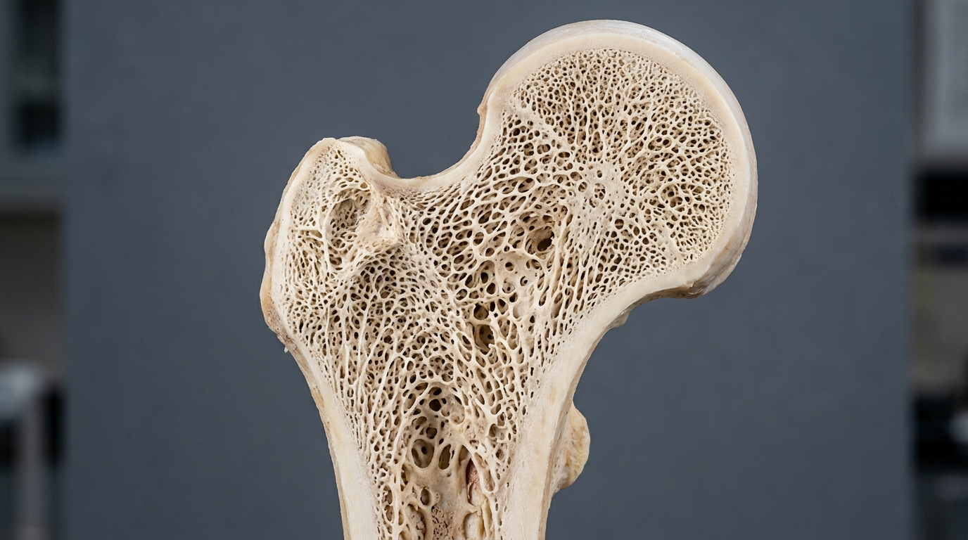
The depth-resolved measurements revealed a consistent structural gradient. Trabecular volumetric tissue mineral density, bone volume to tissue volume, and trabecular thickness all showed an initial decrease in the superficial trabecular regions, followed by a progressive increase toward deeper regions. Cyst-like void volume behaved in the opposite direction, continuously decreasing with depth and almost completely disappearing beyond 9.84 millimeters. In other words, the outer shell of trabecular bone beneath the joint surface is structurally distinct from the deeper core, with lower density, thinner struts, and more void-like spaces near the surface.
Against this backdrop, the statistical analysis separated the two disease signals. Higher OARSI cartilage degeneration grades were associated with higher subchondral mineralized plate thickness, higher trabecular volumetric tissue mineral density, higher bone volume fraction, and thicker trabeculae, but only within the subchondral plate and the superficial to intermediate trabecular zones reaching 13.12 millimeters deep. This pattern is consistent with the well-described phenomenon of subchondral bone sclerosis accompanying cartilage loss, yet the study adds a crucial qualifier: that local remodeling signal fades out with depth and does not reach the innermost region.
Systemic BMD told a different story. The DXA-derived T-score independently predicted trabecular bone parameters at every depth measured, and in the deepest trabecular region, beyond 13.12 millimeters, it was the sole determinant of bone volume to tissue volume and trabecular thickness. Cartilage degeneration showed no association with trabecular bone parameters in that deepest zone. The authors conclude that depth is a critical determinant in the analysis of osteochondral alterations in hip osteoarthritis, and that the two influences on bone structure are spatially distinct rather than globally entangled.
The practical implications follow directly from this spatial map. The authors emphasize that systemic bone mineral density should be treated as a covariate in osteoarthritis research, since it independently shapes trabecular architecture even in specimens obtained from osteoarthritic joints. Conversely, they recommend that deep regions of the femoral head be used for osteoporosis research, because sampling there minimizes potential confounding by local osteoarthritis-related effects. A study of osteoporotic bone quality that analyzes only superficial trabecular bone from an osteoarthritic femoral head risks attributing to systemic bone loss what is actually local joint-driven remodeling, or the reverse.
As an ex vivo study of surgically removed femoral heads from patients with hip osteoarthritis, the work defines how local cartilage degeneration and systemic skeletal status contribute to bone microarchitectural changes at different depths rather than establishing interventions or outcomes in living patients. Its cohort of 30 patients, though spanning the BMD spectrum, represents a single joint site and a single disease population, and the published version is an early-release accepted manuscript subject to further edits. Even so, the depth-resolved framework offers both osteoarthritis and osteoporosis researchers a clearer answer to a deceptively simple question: when bone in the femoral head changes, which disease is speaking, and from how deep.
Subject of Research: Depth-specific bone microarchitecture in the femoral head in relation to osteoarthritis and osteoporosis
Article Title: Depth-specific characterization of bone microarchitecture in the femoral head: implications for osteoarthritis and osteoporosis research
Article References: Simon, A., Alimy, A.-R., Krug, J., Busse, B., Beil, F. T., Amling, M., von Brackel, F. N., & Rolvien, T. (2026). Depth-specific characterization of bone microarchitecture in the femoral head: implications for osteoarthritis and osteoporosis research. Arthritis Research & Therapy, 28(1), Article 194. https://doi.org/10.1186/s13075-026-03943-2
Image Credits: AI Generated
DOI: 10.1186/s13075-026-03943-2
Keywords: femoral head, bone microarchitecture, osteoarthritis, osteoporosis, HR-pQCT, subchondral bone, cartilage degeneration, bone mineral density, DXA, total hip arthroplasty, trabecular bone, OARSI grade
News Source: Ophelia Keating. (November 6, 2026). Bone Depth Map of the Femoral Head Separates Osteoarthritis Effects from Osteoporosis Signals. Scienmag.