A deep learning model trained on routine diagnostic biopsy images can accurately predict whether patients with esophageal adenocarcinoma will achieve a pathological response to preoperative radiochemotherapy under the CROSS protocol, according to research published in Discover Artificial Intelligence. Investigators from the University Hospital of Cologne trained a transformer encoder neural network on 312 whole-slide images of hematoxylin and eosin stained primary tumor biopsies collected from 193 patients with locally advanced esophageal adenocarcinoma who subsequently underwent neoadjuvant chemoradiation followed by surgical resection. In an independent test cohort of 39 patients, the artificial intelligence system achieved an area under the receiver operating characteristic curve of 0.80, successfully discriminating between treatment responders and non-responders prior to the initiation of multimodal therapy. The model demonstrated a sensitivity of 70.59 percent, a specificity of 90.91 percent, a precision of 85.71 percent, and a negative predictive value of 80 percent, with comparable diagnostic performance documented in a separate validation cohort of 31 patients that reached an area under the curve of 0.82.
The study directly addresses a critical clinical challenge in the personalized management of locally advanced esophageal adenocarcinoma, an aggressive malignancy with a rising global incidence that requires intensive multimodal intervention. Standard treatment strategies have traditionally relied on broad evidence-based findings from prospective randomized controlled trials such as ESOPEC, which established perioperative chemotherapy with fluorouracil, leucovorin, oxaliplatin, and docetaxel as an overall standard of care over preoperative radiochemotherapy. More recently, the phase III MATTERHORN trial advanced this paradigm by demonstrating survival advantages through the addition of durvalumab to perioperative chemotherapy. However, population-level trial standards do not ensure therapeutic efficacy for every individual patient, as fewer than 50 percent of patients achieve a favorable pathological response under chemotherapy regimens. Because neoadjuvant radiochemotherapy following the CROSS protocol remains only slightly less effective across broader populations while inducing marked tumor regression in specific patient subsets, an accurate diagnostic prediction tool could identify patients who benefit substantially from preoperative radiation and concurrent carboplatin and paclitaxel.
To evaluate patient responses following neoadjuvant radiochemotherapy and subsequent surgical resection, the researchers utilized the Cologne tumor regression grade system based on the percentage of residual viable tumor cells identified in surgical specimens. Within this grading framework, patients were categorized into four distinct pathological tiers: grade IV representing complete pathological regression with no viable tumor cells, grade III representing major pathological regression with fewer than 10 percent viable tumor cells, grade II representing partial regression with 10 to 50 percent viable tumor cells, and grade I representing no tumor regression with greater than 50 percent viable tumor cells. Because complete and major pathological responses are clinically associated with significant improvements in overall survival compared to minor regression or absent response, the study combined grades III and IV into a single responder group while designating grades I and II as non-responders. Although restricting the positive endpoint exclusively to complete responders would carry clinical appeal in the context of emerging chemoimmunotherapy options, doing so would have produced a severely imbalanced dataset, compromising neural network training and stability.
The patient cohort evaluated in the study consisted of 193 individuals with esophageal adenocarcinoma treated at the University Hospital of Cologne, comprising 88 percent male and 12 percent female patients with a median age of 63 years and a median body mass index of 26.5 kilograms per square meter. Surgical treatment primarily involved hybrid minimally invasive Ivor Lewis esophagectomies combining laparoscopic abdominal mobilization with open thoracotomy in 76 percent of cases, with postoperative complications ranging from none in 35 percent of patients to Clavien-Dindo grade IV in 6.6 percent and grade V mortality in 2.2 percent. Pathological tumor staging of resected specimens identified stage ypT0 in 17 percent, ypT1 in 20 percent, ypT2 in 17 percent, and ypT3 in 46 percent of patients, while regional lymph node involvement was absent in 55 percent, with ypN1 in 20 percent, ypN2 in 17 percent, and ypN3 in 6.4 percent alongside a median resected node count of 31. Assessment of regression grades showed complete response in 15 percent of patients, major response with fewer than 10 percent viable tumor cells in 28 percent, partial response in 38 percent, and no regression in 19 percent, yielding an overall patient-level split of 83 responders across 114 slides and 113 non-responders across 198 slides.
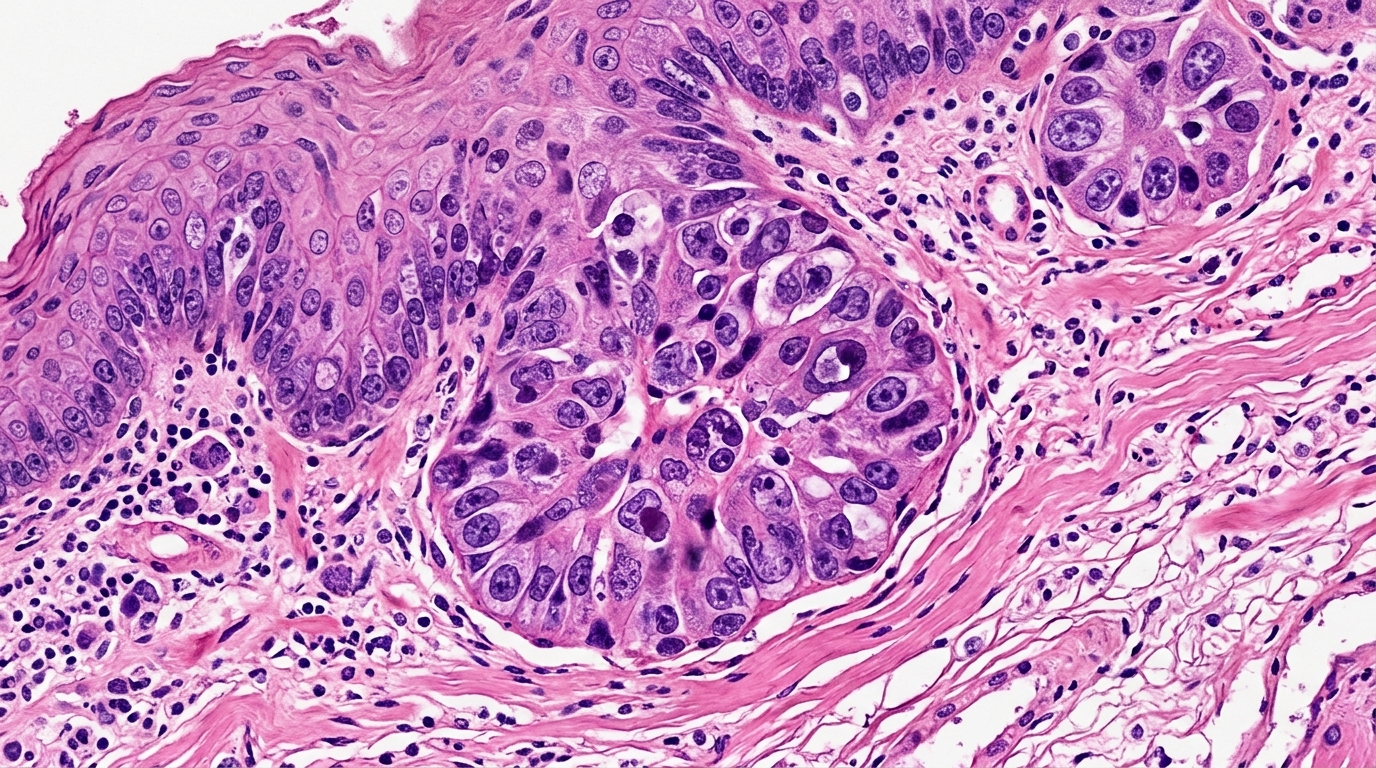
The digitized image analysis pipeline commenced with standardized hematoxylin and eosin staining performed centrally at the University Hospital of Cologne on diagnostic biopsy tissue blocks retrieved from primary diagnostic centers. Sections cut from paraffin-embedded biopsies were digitized at 20x magnification using a Hamamatsu NanoZoomer S360 slide scanner, producing whole-slide images that were subsequently tiled into patches measuring 256 by 256 pixels. Slide background regions lacking tissue were eliminated, leaving an average of 1369 tissue-containing patches from an overall average of 23989 total patches per slide in the test set. Feature vector representations for each valid image patch were generated using the pretrained CTransPath model, representing each whole slide as a unified set of patch-level embedding vectors. When individual patients had multiple biopsy slides, all patch embeddings were merged into a single comprehensive patient profile. These aggregated representations were processed through a transformer encoder whose final layer embeddings were averaged and passed to a linear classification layer, optimized over two training epochs using the Adam optimizer with a learning rate of 0.00025, weight decay of 0.00005, and batch size of 16.
To confirm that the machine learning system learned biological features specific to radiochemotherapy rather than generic markers of treatment sensitivity, the researchers evaluated the trained model on an external cohort of 79 biopsy images from patients treated with the FLOT chemotherapy protocol. When applied to this independent chemotherapy cohort, the network accurately identified only 20 percent of responders, accompanied by a precision of 80 percent. This substantial drop in sensitivity on FLOT-treated tumors indicates that the model is selectively tuned to the distinct biological mechanisms governing response to the CROSS radiochemotherapy regimen, rather than general chemotherapy vulnerability. This divergence underscores that distinct therapeutic modalities are mediated by different tumor microenvironmental factors, highlighting the necessity of establishing separate, dedicated artificial intelligence models tailored specifically to perioperative chemotherapy protocols rather than relying on a single generalized predictive architecture across disparate clinical regimens.
Beyond binary classification, the investigators developed a comprehensive interpretability and explainability framework to identify specific histomorphological and cellular structures driving the neural network decisions. Using Integrated Gradients, the researchers assigned positive attribution scores to whole-slide image patches contributing toward a correct response prediction and negative attribution scores to patches directing the classifier toward non-response. To avoid disproportionate influence from larger tumor specimens, analysis was restricted to the top 10 percent highest-scoring Integrated Gradients patches within manually annotated tumor regions, capped at a maximum of 200 patches per slide. Within these critical regions, cell nuclei were segmented and classified into six distinct populations—epithelial tumor cells, connective cells, lymphocytes, plasma cells, eosinophils, and neutrophils—using a StarDist nuclear segmentation model fine-tuned on the Lizard colorectal histology dataset, with all nuclear classifications independently verified by experienced pathologists.
From the segmented nuclear data, the authors engineered 524 quantitative features describing cell population proportions, nuclear morphology metrics, intercellular distances, and spatial arrangements evaluated via the Common Language Effect Size. Spatial distribution was analyzed using the Average Nearest Neighbor Ratio to measure nuclear dispersion versus clustering, alongside Join Count spatial autocorrelation based on Voronoi tessellation to quantify whether adjacent cells belonged to identical or contrasting cell types. The analysis revealed that patches predictive of radiochemotherapy response contained a significantly higher proportion of tumor cells accompanied by fewer lymphocytes and connective cells, with increased tumor-tumor interactions indicating tight spatial clustering. Conversely, patches predictive of non-response exhibited dispersed tumor cells that interacted extensively with surrounding stromal and immune cells. Notably, total tumor cell counts did not differ between responder and non-responder patches, indicating that spatial clustering and architecture rather than absolute cellular abundance dictate therapeutic sensitivity.
The study authors emphasize that endoscopic biopsies capture only localized fragments of primary tumors, representing an intrinsic limitation given the known spatial intratumoral heterogeneity of gastroesophageal adenocarcinomas. Because single diagnostic biopsies might underrepresent critical morphology, future investigations comparing multiple biopsies from individual tumors will be necessary to evaluate predictive reproducibility in heterogeneous tissue. Nevertheless, because analyzing standard diagnostic biopsies mirrors actual clinical workflows, the investigators have initiated preparations for a prospective validation study supported by funding from the German National Center for Tumor Diseases Investigator-Initiated Trial Portfolio across six additional medical centers. If prospective multi-center validation confirms these diagnostic performance metrics, this computational approach could provide an accessible, cost-effective digital pathology biomarker that identifies patients best suited for individualized CROSS radiochemotherapy, avoiding expensive sequencing while optimizing personalized treatment selection.
Subject of Research: Deep learning prediction of radiochemotherapy response in esophageal adenocarcinoma
Article Title: Artificial intelligence predicts pathological response to neoadjuvant radiochemotherapy from diagnostic biopsies in esophageal adenocarcinoma
Article References: Pisula, J. I., Jung, J.-O., Tolkach, Y., Lyu, S. I., Knipper, K., Damanakis, A., Quaas, A., Bruns, C., Schmidt, T., Bozek, K., & Popp, F. C. (2026). Artificial intelligence predicts pathological response to neoadjuvant radiochemotherapy from diagnostic biopsies in esophageal adenocarcinoma. Discover Artificial Intelligence, 6(1), Article 1790. https://doi.org/10.1007/s44163-026-02411-9
Image Credits: AI Generated
DOI: 10.1007/s44163-026-02411-9
Keywords: Artificial Intelligence, Esophageal Adenocarcinoma, Radiochemotherapy, CROSS Regimen, Digital Pathology, Computational Histopathology, Predictive Oncology, Deep Learning, Artificial, intelligence, predicts, pathological
News Source: Nathaniel Bowman. (November 6, 2026). AI Biopsy Model Predicts Esophageal Cancer Response to Radiochemotherapy. Scienmag.