The ability of lung tissue to repair itself depends on a conversation between progenitor cells and the fibroblasts that surround them, and a new study suggests that conversation changes with age. Researchers working with aged mice and a mouse model of accelerated aging report that lung epithelial progenitor cells retain their capacity to form three-dimensional organoids even in old age, but that the supporting fibroblast niche can undermine this regenerative potential. The work, published as an original research article in Frontiers in Aging, examines how microenvironmental signals shape cell-cell interactions that are essential for lung regeneration.
To probe these changes, the team used wild-type mice aged 18 to 24 months, alongside Zmpste24-deficient mice, which display an accelerated aging phenotype. The researchers assessed regenerative capacity in vitro using a three-dimensional organoid culture system, and characterized isolated cells with markers of senescence and autophagy.
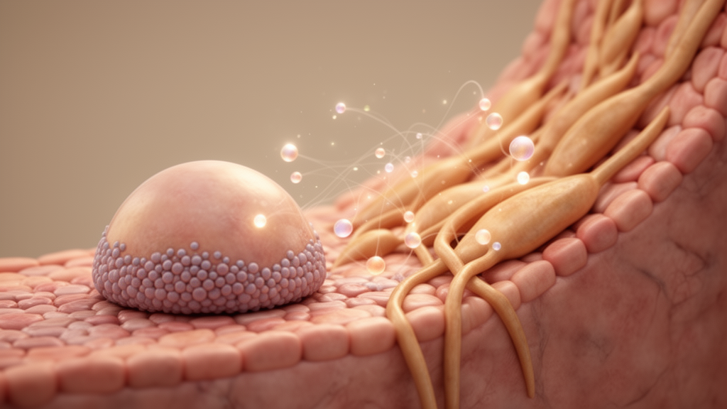
The central finding is that lung epithelial cells from both aged wild-type mice and Zmpste24-deficient mice keep their organoid-forming potential. These progenitors maintained their phenotype and cellular homeostasis through an increased autophagy-associated transcriptional response, an effect that was particularly evident when the epithelial cells were co-cultured with fibroblasts taken from young animals. In other words, the age of the niche matters: young fibroblasts appear to support the compensatory autophagy program that aged progenitors rely on.
The study also points to a mechanical dimension of progenitor aging. The authors report that these lung progenitor cells may become more susceptible to mechanical stress as animals age, potentially in connection with reduced nuclear lamins and the Zmpste24 defect. This vulnerability was illustrated by fluorescence-activated cell sorting, a standard laboratory technique that physically processes cells through sorting equipment: in the study, FACS sorting could further compromise the regenerative potential of the aged progenitor cells. The observation is a caution for experimental workflows, since the very act of isolating cells may itself stress aged, mechanically fragile cells and distort measurements of their true capacity.
The fibroblast side of the story is strikingly different. Cultured fibroblasts from Zmpste24-deficient mice showed nuclear defects and acquired a senescent phenotype, characterized by activation of mechanistic target of rapamycin complex 1, or mTORC1, together with reduced autophagy. This senescent state, in turn, impaired organoid formation. Where aged epithelial cells ramped up autophagy to preserve themselves, the aged-model fibroblasts did the opposite, combining mTORC1 activation with dampened autophagy and losing their ability to support regeneration in culture.
Taken together, the results indicate that in aging, progenitor cells and their fibroblast niche integrate microenvironmental signals that shape the cell-cell interactions essential for lung regeneration. The progenitor compartment appears comparatively resilient, holding on to organoid-forming capacity and compensating transcriptionally, while the niche deteriorates in ways that can be reproduced in a dish. Because the entire study was conducted in mice and in vitro organoid cultures, the findings describe mechanisms in model systems rather than demonstrated effects in human lungs, and the authors note that the impact of aging on adult lung progenitors remains incompletely understood.
The work nonetheless offers a framework for thinking about tissue aging as a two-cell problem rather than a single-cell one. Regenerative failure in an aged organ may reflect not only intrinsic changes in stem cells but also shifts in the supporting cells that provide mechanical and metabolic cues. In this mouse model, the fibroblast niche, marked by nuclear defects, senescence, mTORC1 activation and reduced autophagy, emerges as a decisive partner whose state can tip organoid formation toward success or failure. Disentangling those signals, the authors suggest, is central to understanding how the aged lung regenerates, and why that capacity changes over a lifetime.
Subject of Research: How aging alters interactions between lung epithelial progenitor cells and their fibroblast niche in mouse organoid models of regeneration.
Article Title: Aging remodels progenitor and niche interactions in lung regeneration
Article References: Toscano-Marquez, F., Garcia-Vicente, A., Camacho-Silverio, U., Valdivia-Herrera, T., Río de la Loza, M., Hernández-Xóchihua, E., Ramírez, R., Selman, M., Pardo, A., & Romero, Y. (2026). Aging remodels progenitor and niche interactions in lung regeneration. Frontiers in Aging, 7, Article 1986986. https://doi.org/10.3389/fragi.2026.1986986
Image Credits: AI Generated
DOI: 10.3389/fragi.2026.1986986
Keywords: lung regeneration, progenitor cells, fibroblasts, organoids, aging, autophagy, mTORC1, senescence, Zmpste24, nuclear lamins, mechanical stress, mice
News Source: Beatrice Stafford. (October 28, 2026). Aging Reshapes How Lung Stem Cells and Their Niche Talk to Each Other. Scienmag.