A single-cell RNA sequencing analysis of 38,678 brain cells from an established mouse model of phenylketonuria has identified widespread, cell-type-specific transcriptomic alterations across major brain cell populations. The investigation provides the first comprehensive cellular and molecular map of the brain affected by the condition, capturing distinct gene expression shifts across multiple glial and vascular cell types.
Phenylketonuria is the most common inherited metabolic disorder and is caused by mutations in the PAH gene that trigger hyperphenylalaninemia and brain injury. Although low-phenylalanine dietary therapy alleviates neurological symptoms, the precise cellular and molecular mechanisms driving brain dysfunction have remained unclear, prompting researchers to profile individual brain cells in their established mouse model.
Among the cell populations analyzed, oligodendrocytes exhibited decreased expression of key genes associated with myelination and cellular differentiation. The authors report that these downregulated expression profiles implicate impaired myelination as a key cellular component of phenylketonuria-associated brain injury.
Astrocytes showed a contrasting transcriptional response characterized by increased expression of synapse-organization genes alongside decreased expression of glial-differentiation genes. According to the study, this pattern points to altered astrocytic synaptic support within the injured brain tissue.
The single-cell data also revealed metabolic and neurovascular disturbances in other cell types. Microglia displayed upregulation of genes involved in mitochondrial oxidative phosphorylation, indicating microglial metabolic activation, while endothelial cells showed bidirectional dysregulation of genes regulating vascular homeostasis and remodeling, reflecting neurovascular dysfunction.
The investigators conclude that these coordinated transcriptomic changes reveal how impaired myelination, altered astrocytic synaptic support, microglial metabolic activation, and neurovascular dysfunction converge in phenylketonuria-associated brain damage. The resulting cellular map provides new insight into the disorder’s complex pathophysiology to help guide the future development of specific therapeutics beyond dietary management.
Subject of Research: Single-cell transcriptomic profiling of brain tissue in a phenylketonuria mouse model
Article Title: Single-cell RNA sequencing of brain tissue from a mouse model of phenylketonuria
Article References: Gao, H., Liu, W., Zhang, C., Jin, X., Zhong, W., Zhai, Y., Yao, Z., Yan, H., Hao, W., Wang, Y., Cao, X., Cao, Z., Yan, Y., & Yin, C. (2026). Single-cell RNA sequencing of brain tissue from a mouse model of phenylketonuria. Orphanet Journal of Rare Diseases. https://doi.org/10.1186/s13023-026-04613-x
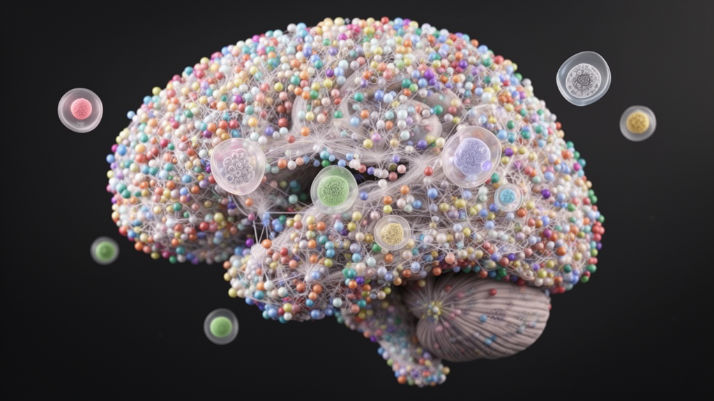
Image Credits: AI Generated
DOI: 10.1186/s13023-026-04613-x
Keywords: phenylketonuria, single-cell RNA sequencing, PAH mutations, hyperphenylalaninemia, brain dysfunction, oligodendrocytes, myelination, astrocytes, synapse organization, microglia, mitochondrial oxidative phosphorylation, endothelial cells, vascular homeostasis, neurovascular dysfunction, mouse model, transcriptomic alterations, glial differentiation
News Source: Cassandra Pierce. (November 16, 2026). Single-Cell Sequencing Maps Diverse Brain Cell Alterations in Phenylketonuria Mouse Model. Scienmag.