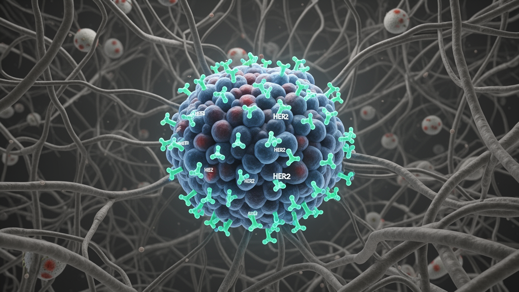
A comprehensive scientific review published in Experimental Hematology and Oncology synthesizes the biological mechanisms, tumor microenvironment features, diagnostic challenges, and translational treatment strategies governing human epidermal growth factor receptor 2, known as HER2, across diverse solid tumors. Encoded by the ERBB2 gene, HER2 belongs to the epidermal growth factor receptor family and functions as an established oncogenic driver most prominently identified in breast and gastric cancers. Unlike other epidermal growth factor receptor family members, the HER2 receptor protein lacks a canonical ligand-binding domain and instead exists in a constitutively active structural conformation that promotes spontaneous receptor dimerization and intracellular signal amplification. Acting as a pivotal molecular switch in neoplastic development, HER2 stimulates multiple intracellular cascades, including the Ras, Raf, and mitogen-activated protein kinase pathway, the phosphatidylinositol 3-kinase and Akt pathway, the signal transducer and activator of transcription pathway, and the phospholipase C cascade. The synthesis highlights how these downstream cascades coordinate cellular proliferation, survival, invasive behavior, and therapeutic responsiveness across distinct neoplastic settings.
Expression and functional activation of HER2 are governed by multi-tiered regulatory mechanisms operating at transcriptional, epigenetic, and post-translational tiers. Transcriptional regulators including Sp1, AP-2, and Ets-1, alongside epigenetic remodeling and non-coding ribonucleic acids, modulate baseline gene readout, while subsequent post-translational modifications like phosphorylation, ubiquitination, and glycosylation control the final stability, subcellular localization, and enzymatic activity of the receptor. Beyond intrinsic intracellular signaling, HER2 functions as an immunogenic tumor-associated antigen whose surface overexpression allows major histocompatibility complex class II molecules to process and present derived peptides, stimulating helper CD4 positive T lymphocyte responses. Simultaneously, HER2 signaling actively dampens host antitumor immunity by interfering with intracellular cGAS-STING signaling, shielding malignant cells from immunological surveillance. HER2 activation triggers cellular interleukin-6 secretion and downstream STAT3 signaling, which suppresses antitumor T-cell activity by averting apoptosis and preventing naive CD4 positive T cells from differentiating into regulatory T cells. Cancer cells also maintain interleukin-8 secretion, up-regulating programmed cell death protein 1 on CD8 positive T cells and fostering local immunosuppression.
The tumor microenvironment surrounding HER2-altered neoplasms represents a dynamic ecosystem shaped through bidirectional signaling between malignant cells, infiltrating immune populations, and surrounding stromal elements. HER2-positive cancer cells recruit B lymphocytes into the microenvironment by secreting chemokines including CCL19, CCL21, and CXCL13, and high baseline B-cell infiltration correlates with favorable clinical prognosis. Simultaneously, HER2-driven phosphatidylinositol 3-kinase, Akt, and mitogen-activated protein kinase signaling induces macrophage polarization toward an immunosuppressive M2 phenotype. In response, tumor-associated macrophages secrete growth factors including vascular endothelial growth factor and transforming growth factor-beta, as well as interleukins 6 and 10, which feed back to sustain HER2 receptor signaling. Interaction with adjacent stromal cells further enhances disease progression, as HER2 activation stimulates matrix metalloproteinases that degrade extracellular matrix barriers while upregulating hypoxia-inducible factor 1-alpha to drive vascular endothelial growth factor-mediated angiogenesis. In parallel, surrounding cancer-associated fibroblasts release cytokines and growth factors like basic fibroblast growth factor and hepatocyte growth factor to stimulate HER2 activity, whereas therapeutic inhibition of epidermal growth factor receptor and HER2 signaling specifically depletes these supporting fibroblasts.
Genomic alterations involving HER2 exhibit substantial structural diversity across solid malignancies, encompassing gene amplification, somatic base mutations, and varied protein expression levels. Gene amplification elevates ERBB2 copy numbers, thereby driving marked receptor overexpression on the cellular membrane, although protein overexpression can also occur without amplification due to aberrant upstream pathway activation. Evolutionary analyses demonstrate that HER2 amplification is a late-stage genomic event that remains mutually exclusive with early RAS and RAF driver mutations, emerging to adapt tumor cells to microenvironmental niches and promote invasive metastasis. When RAS and RAF mutations have already fully activated the mitogen-activated protein kinase cascade, additional HER2 signaling becomes redundant or cytotoxic, leading to growth inhibition or apoptosis. In contrast, somatic ERBB2 mutations cluster within the intracellular tyrosine kinase domain, altering kinase activity and surface presentation. These mutations exhibit notable tumor specificity, such as exon 20 insertions like Y772_A775dup and G776delinsVC in non-small cell lung cancer, widespread mutations exceeding thirty percent in urothelial carcinoma, and kinase-domain substitutions like L755S, R784G, and V842I across colorectal and gastric malignancies.
Clinical management of HER2-driven cancers is complicated by pronounced spatial and temporal heterogeneity across primary and metastatic lesions. Spatial interlesional and intratumoral heterogeneity presents distinct architectural patterns, described in breast tumors as clustered, mosaic, or dispersed distributions of amplified cells. Gastric cancers display especially high rates of spatial variation, with immunohistochemical heterogeneity identified in forty-five to seventy-nine percent of patients and gene amplification heterogeneity detected in twenty-three to fifty-four percent of cases. Dual-assay evaluations utilizing the gene-protein assay method differentiate between genetic heterogeneity, where amplified and non-amplified clones coexist, and non-genetic heterogeneity, where cells harbor gene amplification without corresponding protein overexpression. Temporally, receptor expression fluctuates across serial disease stages and treatment courses; for example, forty-two point eight percent of breast tumors classified as non-expressing on initial core biopsy demonstrate low expression upon surgical resection, while twenty-nine point one percent of first-line trastuzumab-treated gastric cancer patients in the GASTHER3 study experienced complete HER2 loss. Standardized endoscopic sampling of six to eight tissue fragments and dual-block testing methods are employed to reduce diagnostic misclassification.
To overcome diagnostic discrepancies arising from dynamic tumor heterogeneity, pathological evaluation has expanded to incorporate liquid biopsy platforms and artificial intelligence algorithms. Minimally invasive liquid biopsy approaches isolate circulating tumor cells and cell-free circulating tumor DNA from peripheral blood samples, allowing longitudinal assessment of ERBB2 amplification using digital polymerase chain reaction and droplet digital techniques. Clinical investigations in non-breast, non-gastric solid tumors identified ERBB2 amplification in baseline plasma from eighty-five percent of evaluated patients, establishing a significant positive correlation with formal tissue sequencing. Concurrently, digital image analysis and artificial intelligence models evaluate whole-slide immunohistochemical imaging by quantifying staining intensity, membrane completeness, and positive cell percentages. Independent studies confirm that algorithmic assistance elevates diagnostic agreement among reviewing pathologists from forty-nine point three percent to seventy-four point one percent, significantly refining the classification of ambiguous low-expressing cases. Furthermore, multimodal deep-learning frameworks integrating pretreatment mammography, ultrasound, and magnetic resonance imaging achieve predictive receiver operating characteristic area under curve values of 0.836 for receptor status and 0.858 for neoadjuvant chemotherapy response.
The therapeutic paradigm for HER2-driven solid tumors has evolved from foundational single-agent regimens to rational combination strategies and engineered pharmacological modalities. Clinically approved therapies encompass monoclonal antibodies, small-molecule tyrosine kinase inhibitors, and antibody-drug conjugates. Monoclonal antibodies like trastuzumab and pertuzumab bind extracellular domains IV and II, respectively, blocking receptor dimerization, inhibiting downstream signaling cascades, and triggering antibody-dependent cellular cytotoxicity. Second-generation monoclonal agents like margetuximab incorporate engineered fragment crystallizable regions to elevate immune effector cell engagement. Small-molecule tyrosine kinase inhibitors, including lapatinib, neratinib, pyrotinib, poziotinib, and tucatinib, reversibly or irreversibly block intracellular phosphorylation and penetrate the blood-brain barrier to control intracranial metastases, though older agents frequently induce toxicities through off-target epidermal growth factor receptor inhibition. Newer selective oral inhibitors like zongertinib selectively spare wild-type epidermal growth factor receptors to minimize adverse events, whereas sevabertinib reversibly targets challenging exon 20 insertion mutations. Novel bispecific constructs like zanidatamab simultaneously engage distinct non-overlapping epitopes, gaining clinical approvals in advanced HER2-positive biliary tract cancers.
Antibody-drug conjugates represent an impactful translational class by coupling targeted monoclonal antibodies to potent cytotoxic payloads using specialized chemical linkers. Conjugate efficacy depends on the drug-to-antibody ratio, which typically spans between two and eight payload molecules per antibody scaffold. Modern conjugates such as trastuzumab deruxtecan and disitamab vedotin demonstrate superior clinical activity over earlier agents like trastuzumab emtansine by leveraging membrane-permeable cleaved toxins to execute bystander killing, effectively eliminating adjacent low-expressing or antigen-negative cells within heterogeneous tumors. In the Chinese pharmaceutical pipeline, disitamab vedotin incorporates monomethyl auristatin E to target low-expression settings, while SHR-A1811 introduces the potent payload rezatapotecan along with a stabilizing chiral cyclopropyl linker. Concurrently, combining HER2-targeted agents with immune checkpoint inhibitors exhibits varying efficacy across solid tumors. While the phase III KEYNOTE-811 trial demonstrated that adding pembrolizumab to trastuzumab and chemotherapy prolonged survival in advanced gastric cancer with combined positive score of at least one, combination trials in non-small cell lung cancer like DESTINY-Lung03 produced serious adverse event rates of sixty-three point six percent without survival advantages over monotherapy.
Tumor microenvironment landscapes differ considerably across anatomical cancer types, dictating therapeutic sensitivity and patterns of acquired resistance. In breast cancer, high baseline tumor-infiltrating lymphocyte density correlates with improved pathologic complete response rates, whereas immunosuppressive components like M2 macrophages and programmed death-ligand 1 expression modulate clinical responsiveness. Resistance mechanisms involve podoplanin-positive cancer-associated fibroblasts that secrete indoleamine 2,3-dioxygenase 1 and tryptophan 2,3-dioxygenase 2 to suppress antibody-dependent cytotoxicity, as well as angiogenic vascular endothelial growth factor overexpression. In gastric cancer, HER2-overexpressing regions show decreased CD8 positive T-cell densities and diminished STING pathway activity relative to low-expressing areas. Tumors in gastric settings frequently release microvesicles containing glutaminase 1 via CDC42 pathways, driving M2 macrophage polarization and acquired trastuzumab resistance. In non-small cell lung cancer, tumors harboring HER2 exon 20 insertions display cold immunological profiles, low response to immune checkpoint inhibition, and frequent concomitant activation of phosphatidylinositol 3-kinase signaling, whereas HER2-amplified colorectal tumors exhibit elevated tertiary lymphoid structures and high tumor-infiltrating lymphocytes without direct prognostic correlations.
To counteract acquired resistance arising from target loss, pathway compensation, and microenvironmental immunosuppression, investigational oncology is exploring multispecific antibodies, adoptive cellular therapies, and therapeutic cancer vaccines. Multispecific constructs like the trispecific antibody SAR443216 co-engage HER2, CD3, and CD28 to recruit T cells, while DF1001 bridges HER2 with CD16 and NKG2D to activate natural killer and cytotoxic T cells. Adoptive cell therapies under evaluation encompass engineered chimeric antigen receptor T cells, natural killer cells, and macrophages. In a phase I trial of HER2-directed chimeric antigen receptor T cells involving eleven patients with biliary or pancreatic cancers, disease control was achieved in six individuals without severe toxicity, while preclinical engineered universal platforms counter immunosuppressive transforming growth factor-beta signaling. Concurrently, chimeric antigen receptor macrophages like CT-0508 demonstrate safety and local microenvironmental remodeling in phase I trials. Complementary therapeutic vaccines, such as intracellular domain deoxyribonucleic acid vaccines, dendritic cell platforms achieving seventy-two point seven percent pathologic complete response in early-stage breast cancer, and the peptide vaccine HER-Vaxx prolonging gastric cancer survival, illustrate an expanding precision armamentarium targeting HER2-altered malignancies.
Subject of Research: Tumor microenvironment biology, spatiotemporal heterogeneity, and translational therapeutic strategies in HER2-altered solid tumors
Article Title: HER2 in solid tumors: microenvironment features and multifaceted advances from mechanisms to clinical translation
Article References: Zhang, T., Tan, C., Sun, H., Gu, W., Wang, W., Xu, M., & Sheng, W. (2026). HER2 in solid tumors: microenvironment features and multifaceted advances from mechanisms to clinical translation. Experimental Hematology & Oncology, 15(1), Article 114. https://doi.org/10.1186/s40164-026-00850-9
Image Credits: AI Generated
DOI: 10.1186/s40164-026-00850-9
Keywords: HER2, Solid Tumors, Tumor Microenvironment, Cancer Heterogeneity, Antibody-Drug Conjugates, Liquid Biopsy, Artificial Intelligence, Immunotherapy, Trastuzumab Deruxtecan, Disitamab Vedotin, Tyrosine Kinase Inhibitors, Bispecific Antibodies, Adoptive Cell Therapy, Cancer Vaccines, Breast Cancer, Gastric Cancer
News Source: Nathaniel Bowman. (November 16, 2026). Review Synthesizes Solid Tumor Microenvironment Dynamics and Emerging HER2 Targeted Strategies. Scienmag.