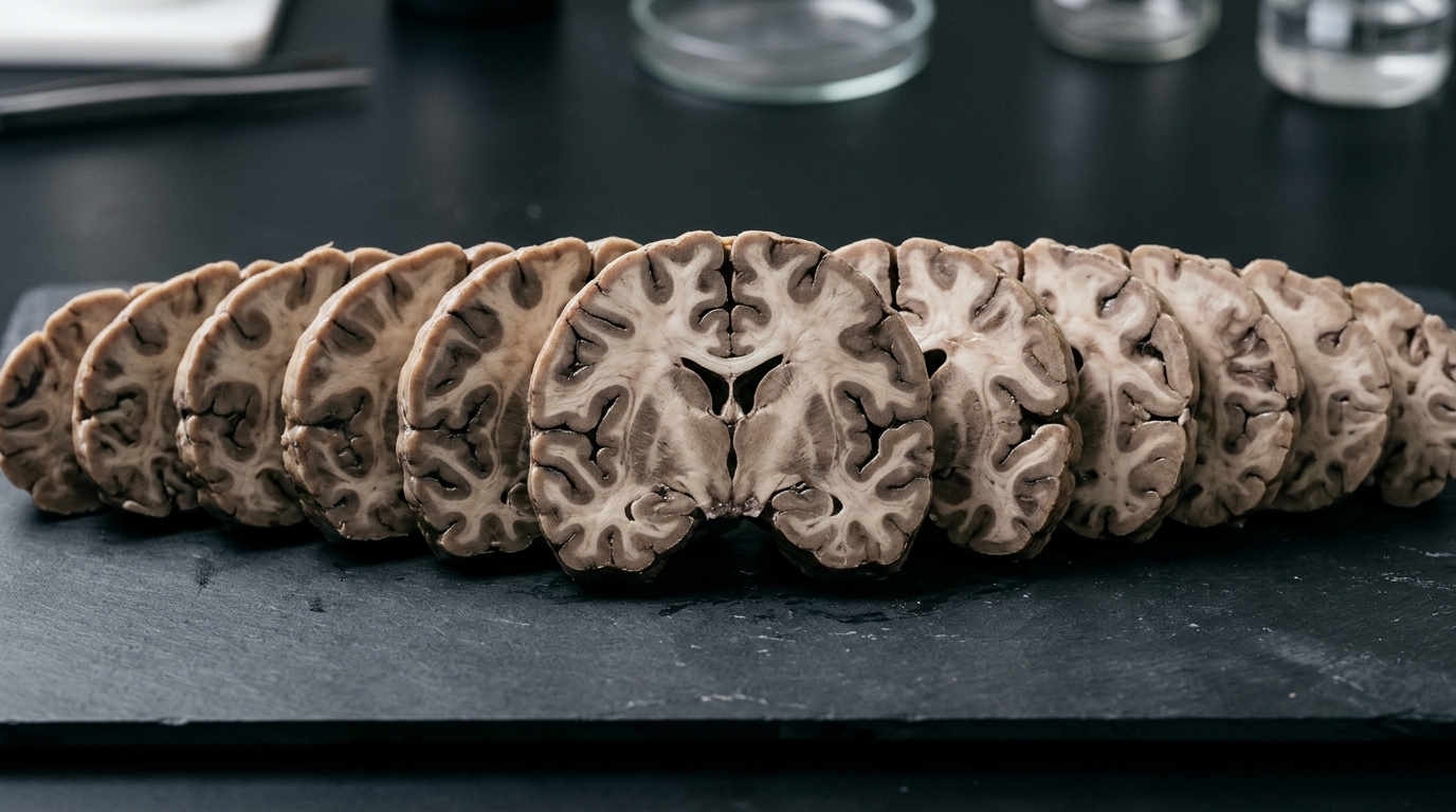
Prior work has shown the feasibility of reconstructing 3D brain volumes from 2D dissection photographs, but these outputs often exhibit coarse, overly smooth reconstructions of structures, especially under high anisotropy. A new study published in Acta Neuropathologica Communications introduces a computationally efficient super-resolution step that imputes slices to generate anatomically consistent isotropic volumes from anisotropic 3D reconstructions of dissection photographs. The approach, developed by researchers including Marina Crespo-Aguirre, Jonathan Williams-Ramirez, Dina Zemlyanker, and senior author Juan Eugenio Iglesias, addresses a key limitation in prior work that reconstructed 3D brain volumes from 2D dissection photographs but yielded coarse, overly smooth renderings of brain structures, especially under high anisotropy—that is, when reconstructing from thick slabs.
The new method tackles this by training on domain-randomized synthetic data, a strategy the authors say ensures the technique generalizes across dissection protocols and remains robust to large slab thicknesses and hallucination effects.
The researchers report that the imputed volumes yield improved automated segmentations, achieving higher Dice scores—a standard metric for measuring overlap between automated and reference segmentations—particularly in cortical and white matter regions.
Beyond segmentation, the team validated the approach on two downstream tasks: surface reconstruction and atlas registration. The imputed volumes produced more accurate cortical surfaces, meaning the outer boundary of the brain could be modelled with greater geometric fidelity. Alignment with the target template also improved, indicating that photograph-based reconstructions could be more reliably mapped onto standardized brain atlases.
The broader significance lies in what the authors describe as strengthening the bridge between neuropathology and neuroimaging. Neuropathological analyses benefit from spatially precise volumetric reconstructions that enhance anatomical delineation and improve morphometric accuracy.
The method is computationally efficient, which the authors highlight as an advantage for practical adoption.
The method is publicly available at https://surfer.nmr.mgh.harvard.edu/fswiki/mri_3d_photo_recon, allowing other research groups to adopt and evaluate the technique. The article was published on 12 October 2026 and is open access.
Subject of Research: AI-based slice imputation to improve 3D brain reconstruction fidelity from neuropathology dissection photographs
Article Title: Improving neuropathological reconstruction fidelity via AI slice imputation
Article References: Crespo-Aguirre, M., Williams-Ramirez, J., Zemlyanker, D., Hu, X., Deden-Binder, L. J., Herisse, R., Montine, M., Connors, T. R., Mount, C., MacDonald, C. L., Keene, C. D., Latimer, C. S., Oakley, D. H., Hyman, B. T., Lawry-Aguila, A., & Iglesias, J. E. (2026). Improving neuropathological reconstruction fidelity via AI slice imputation. Acta Neuropathologica Communications. https://doi.org/10.1186/s40478-026-02451-3
Image Credits: AI Generated
DOI: 10.1186/s40478-026-02451-3
Keywords: neuropathology, 3D reconstruction, dissection photographs, super-resolution, slice imputation, anisotropy, automated segmentation, Dice score, cortical surfaces, atlas registration, domain randomization, neuroimaging
News Source: Cassandra Pierce. (November 5, 2026). AI Fills the Gaps: Sharper 3D Brain Reconstructions from Dissection Photographs. Scienmag.