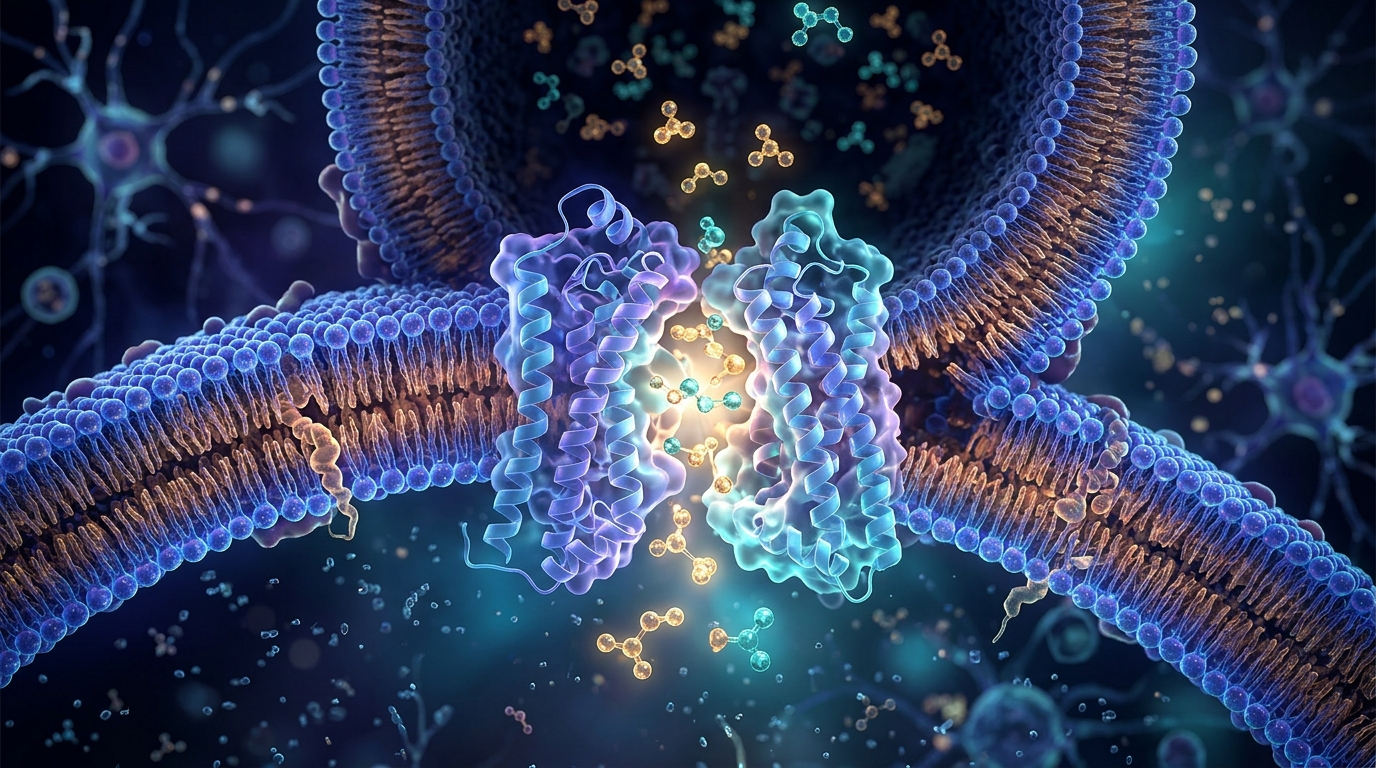
The vesicular inhibitory amino acid transporter, or VIAAT, is the single molecular machine responsible for loading GABA and glycine into synaptic vesicles in the human brain. Without it, inhibitory neurotransmission—the braking system that keeps neural circuits stable and prevents pathological hyperexcitability—would fail entirely. In mice, knocking out the transporter causes embryonic lethality and severe developmental malformations, while in humans, missense mutations in the VIAAT gene impair GABAergic signaling and underlie a spectrum of genetic epilepsies. Despite this central role, two fundamental questions have persisted: how does one transporter recognize two chemically distinct neurotransmitters, and what ionic force actually drives their transport into vesicles? A new study published in Cell Research now provides detailed answers to both, combining high-resolution cryo-electron microscopy, molecular dynamics simulations, and functional assays in cultured neurons.
The research team, led by Yiqing Wei and Yan Zhao, engineered a wild-type human VIAAT construct carrying a C-terminal Twin-Strep tag for purification and an IRES-mCherry cassette to monitor expression. To confirm the construct was physiologically active, they performed whole-cell patch-clamp recordings in cultured hippocampal neurons. When endogenous VIAAT was knocked down with shRNA, inhibitory postsynaptic currents disappeared; when the tagged, shRNA-resistant construct was expressed, synaptic transmission was restored. With this validated system in hand, the researchers determined cryo-EM structures of VIAAT in four states: unbound (apo), GABA-bound, glycine-bound, and apo in chloride-free phosphate buffer, at resolutions of 3.3 Å, 2.7 Å, 3.1 Å, and 3.0 Å respectively.
Human VIAAT comprises 525 amino acids, of which residues 113 through 521 were resolved in the structures. The protein adopts an 11-transmembrane architecture built from two inverted repeats—transmembrane helices 1–5 and 6–10—plus a peripheral helix, TM11, forming what structural biologists call a classical LeuT fold. The apo structure revealed a large cavity open toward the vesicle lumen but closed on the cytosolic side, consistent with a lumen-facing open conformation. Compared with the outward-facing glycine transporter GlyT1, VIAAT retains the canonical SLC6 fold but shows distinct features: its extracellular loop 4 is markedly shorter, leaving the luminal vestibule more exposed and enlarging the space above the substrate-binding pocket. VIAAT also contains a pronounced π-helical bulge in TM10 that shifts the helix toward TM6, and a prominent TM2–TM3 loop on the cytosolic side, enriched in charged residues, that forms a short antiparallel β-sheet.
The structures also offer a framework for interpreting disease-causing mutations. The pathogenic variant F322C maps directly to the substrate-binding pocket and is likely to impair substrate coordination, while P395L and L468P, located in the central regions of TM8 and TM10, are expected to disrupt helix geometry. Three other epilepsy-linked variants—G461D, T464R, and G465S—cluster in TM10, a helix implicated in regulating extracellular gate dynamics, and may impair transport by perturbing that process. Functional analyses confirmed the structural predictions: all six mutants displayed markedly reduced transport activity.
To understand how VIAAT recognizes its two substrates, the team compared the GABA-bound and glycine-bound structures with the apo form. In both complexes, a well-defined rod-shaped density appeared in the central cavity—absent in the apo structure—consistent with the bound substrate. The GABA-bound structure adopts a lumen-facing occluded state, with the binding pocket inaccessible from both the luminal and cytosolic sides. Within this pocket, the carboxyl group of GABA forms hydrogen bonds with the hydroxyl group of Y221 on TM3 and the backbone amides of M132 and F133 on TM1, along with water-mediated interactions involving G131, S316, and K351. The amino group hydrogen-bonds with A128 and T318, and is further stabilized by a water-mediated network including N127, E213, and T217. Glycine adopts a remarkably similar pose, engaging the same key residues through equivalent interactions. Mutagenesis validated these contacts: Y221F, Y221A, and T318A nearly abolished transport activity, while N127A, F133A, and T217A caused moderate reductions. After depletion of inhibitory synaptic vesicles by train stimulation, the N127A and F133A mutants also showed significantly slower recovery of inhibitory postsynaptic currents than the wild type.
A striking structural feature emerged from comparisons with related transporters. The residue F315 on TM6 adopts distinct conformations in the substrate-bound structures: in the GABA-bound state it samples two rotamers, one pointing toward the lumen and TM10 to create a continuous luminal pathway, the other pointing toward TM3 to contribute to GABA binding and seal the cavity in its occluded state. In the glycine-bound structure, F315 predominantly adopts the first rotamer. Substituting F315 with alanine nearly abolished transport, underscoring its critical role in conformational transitions. Perhaps most telling, the binding cavity of VIAAT measures roughly 323 cubic angstroms—far larger than the ~122 cubic angstroms of GlyT1, the ~89 of GlyT2, or the ~178 of GAT1. This enlarged pocket likely explains why VIAAT binds GABA and glycine with relatively low affinity, a property that may suit a transporter that must accommodate two different neurotransmitters.
The question of what drives VIAAT-mediated transport has long been controversial, with previous studies proposing either proton or chloride gradients as the energetic force. The new structures identified two putative chloride-binding sites, designated Cl1 and Cl2, based on characteristic coordination geometry and local density features. The Cl1 site is coordinated by N127, Q130, T271, H274, and K389, while Cl2 is defined by S319, Q320, and H344. Distinct sphere-shaped densities appeared at both positions in the GABA-bound map but were absent in the chloride-free phosphate structure. Molecular dynamics simulations showed persistent chloride occupancy at these sites over 400 nanoseconds, providing additional support for the assignment. Notably, the Cl1 site roughly overlaps with the Na2 sodium-binding site in GlyT1, which stabilizes the outward-facing conformation in LeuT-fold transporters, suggesting the chloride at Cl1 may fulfill a similar structural role. Mutations H274A, S319A, Q320A, and H344A markedly impaired transport activity. The authors caution, however, that the physiological relevance of these sites requires further validation.
The proton side of the coupling question centered on E213, the only titratable residue within the highly electronegative substrate-binding pocket, and one that is highly conserved across VIAAT orthologs. Its predicted pKa in the apo structure is 6.59, compatible with protonation on the luminal side under physiological vesicular pH. A cytosol-facing homology model built on SLC38A9 as a template showed E213 accessible from the cytoplasmic side with a predicted pKa of 6.67, meaning it would be deprotonated at cytosolic pH. These observations suggest E213 can undergo alternating protonation and deprotonation during the transport cycle. Functionally, both E213A and E213Q abolished transport activity, while E213D—which retains a titratable carboxyl group—performed comparably to the wild type. All-atom simulations reinforced the picture: when E213 was protonated, GABA adopted a single dominant conformation closely resembling the crystallographic pose; when E213 was deprotonated, GABA sampled multiple distinct clusters with considerable conformational variation, its amino group displaced into stronger electrostatic contact with E213. Protonation state, in other words, appears to modulate substrate binding and release.
The simulations also disentangled the roles of the two chloride sites. Both remained highly stable throughout each trajectory regardless of E213 protonation state. With E213 protonated, Cl2 stayed stably coordinated even after Cl1 was removed, and GABA kept one predominant pose. In contrast, simulations started from a Cl2-depleted state revealed pronounced instability in GABA binding, with the substrate sampling multiple distinct poses and Cl1 drifting from its initial site. The team concludes that Cl1 is likely not essential for substrate association but instead facilitates conformational transitions, analogous to the Na2 site in other LeuT-fold transporters, whereas Cl2 appears critical for substrate binding itself, since its depletion significantly destabilizes GABA.
Finally, the study mapped the conformational transitions that shuttle VIAAT between its lumen-facing and cytosol-facing states. In the lumen-facing apo structure, TM1a is held close to the scaffold domain by a salt bridge between K113 and E403, and by electrostatic interactions between E120 and K261 and K265—contacts that prevent the dilation of TM1 needed to reach the cytosol-facing conformation. In the cytosol-facing model, a compact electrostatic and hydrogen-bonding network formed by Y139, H143, K305, E368, and H452 stabilizes that state. These residues are not conserved across the SLC6 family but are highly conserved among VIAAT homologs from different species, pointing to a VIAAT-specific gating mechanism. Alanine substitutions of K113, E120, K305, or E368 severely impaired or abolished transport. The authors note an important limitation: conventional whole-cell patch-clamp recordings in VIAAT-expressing HEK293T cells did not yield reliable GABA-evoked currents, so direct measurements of substrate affinity and ion gradient-controlled reconstitution experiments will be needed to fully define the roles of chloride and protons. Even so, the work delivers a structural and mechanistic framework for how the brain’s inhibitory neurotransmitter loader recognizes its substrates, couples ion gradients to transport, and switches between conformational states—a foundation for understanding VIAAT dysfunction in epilepsy and related neurodevelopmental disorders.
Subject of Research: Structural and mechanistic basis of substrate recognition and ion coupling in the human vesicular inhibitory amino acid transporter VIAAT
Article Title: Substrate recognition and ion-coupling mechanism of human VIAAT
Article References: Wei, Y., Ma, K., Hao, K., Bai, Q., Zhao, J., Xie, X., Li, R., Chen, Q., Li, Y., Zhang, H., Cao, Z., Hu, T., & Zhao, Y. (2026). Substrate recognition and ion-coupling mechanism of human VIAAT. Cell Research. https://doi.org/10.1038/s41422-026-01299-5
Image Credits: AI Generated
DOI: 10.1038/s41422-026-01299-5
Keywords: VIAAT, GABA, glycine, cryo-EM, SLC6 transporter, chloride coupling, proton coupling, inhibitory neurotransmission, synaptic vesicles, molecular dynamics, genetic epilepsy, LeuT fold
News Source: Cassandra Pierce. (November 4, 2026). Scientists Solve Structure of the Transporter That Packs GABA and Glycine Into Brain Vesicles. Scienmag.