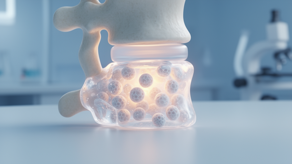
Scientists in China have built an injectable hydrogel seeded with lab-grown cartilage microtissues that preserved disc height and shifted the molecular balance inside degenerated intervertebral discs in an animal model, according to a study accepted for publication in Frontiers in Cell and Developmental Biology. The composite, called CM-Gel, was designed to tackle a problem that current clinical interventions for low back pain rarely solve: the degenerated nucleus pulposus, the gel-like core of the disc, does not effectively regenerate on its own once degeneration has set in.
The research team, led by investigators at Qingdao Haici Hospital, generated cartilage microtissues containing cartilage-specific extracellular matrix through three-dimensional culture in the laboratory. These microtissues were then integrated into a hydrogel to form the injectable composite. The strategy behind the design is straightforward: rather than delivering isolated cells, the material carries pre-formed microtissues that already possess a matrix environment resembling the tissue they are meant to repair.
Before any animal testing, the group tracked how the microtissues matured inside the hydrogel. Using immunofluorescence double staining on days 1, 4, and 7 of in vitro culture, the researchers measured the dynamic expression of two cartilage markers, ACAN and COL-II. Both increased in a time-dependent manner over the seven-day window, which the authors interpret as progressive maturation of the microtissue phenotype. In other words, the longer the microtissues sat in the gel, the more strongly they expressed the molecular signature of healthy cartilage-like tissue.
The in vivo phase used an established animal model of intervertebral disc degeneration. Four weeks after surgery, tissue samples were collected from the treated discs and analyzed. Imaging and histological assessments showed that discs receiving CM-Gel retained their height better than untreated degenerated discs, and the extracellular matrix within the nucleus pulposus showed signs of regeneration. Disc height loss is one of the structural hallmarks of degeneration, so its preservation is a meaningful structural endpoint, though the four-week time frame captures only the early phase of repair.
The molecular readouts told a more detailed story. Quantitative reverse-transcription PCR measured mRNA levels of anabolic genes, catabolic genes, and a fibrosis marker gene in the nucleus pulposus tissue. In the CM-Gel group, the anabolic markers ACAN, COL-II, and SOX9 were significantly upregulated. SOX9 is a transcription factor that drives the expression of matrix genes, so its elevation alongside the structural genes suggests the treated tissue had shifted toward a matrix-building state.
At the same time, the catabolic enzymes MMP13 and ADAMTS5 were significantly downregulated, as was COL-I, the fibrosis marker. MMP13 and ADAMTS5 are enzymes that break down extracellular matrix components, and their reduction indicates suppressed matrix degradation. The drop in COL-I points to less fibrotic remodeling, a process in which the disc’s soft core is progressively replaced by stiffer scar-like tissue. Taken together, the authors argue, these changes show that CM-Gel promotes matrix synthesis, inhibits degradation, and mitigates fibrosis within the treated discs.
The study’s central claim is that the composite reestablishes the balance between synthesis and catabolism, the equilibrium whose disruption defines degenerative disc disease. When catabolic activity outpaces matrix production, the disc loses its structural integrity and its biological function deteriorates. By simultaneously raising anabolic gene expression and lowering catabolic and fibrotic signals, the treatment appears to have moved the degenerative microenvironment back toward a regenerative state in this model.
Several caveats frame these findings. The work is a preclinical animal study with a four-week observation period, and the abstract describes the results as demonstrating potential rather than established clinical benefit. The authors themselves position CM-Gel as a novel strategy with potential for clinical translation, language that signals the distance still separating an animal model result from a therapy for patients with low back pain. Human discs differ from animal discs in size, loading, and degeneration patterns, and longer studies would be needed to assess durability of the structural and molecular changes.
Still, the approach reflects a broader trend in regenerative medicine toward engineered microtissues rather than single-cell suspensions. Because the cartilage microtissues arrive with their own extracellular matrix already in place, they may provide both structural support and a reservoir of matrix signals upon injection. The combination of an injectable format, which allows minimally invasive delivery into the disc space, with a biologically active payload distinguishes this system from purely structural biomaterial implants.
The study was received on 30 June 2026 and accepted on 17 October 2026, and the published version notes that the final formatted article will appear shortly. For the field of intervertebral disc regeneration, the results add to a growing body of evidence that biomaterial-carried microtissues can influence the degenerative microenvironment, at least in the short term and in animal models. Whether that influence translates into durable repair in human spines remains the question the next phase of research will have to answer.
Subject of Research: Preclinical testing of a cartilage microtissue-loaded injectable hydrogel for repairing degenerated intervertebral discs
Article Title: Synchronous restoration of intervertebral disc structure and biological function utilizing cartilage microtissue-based active hydrogels
Article References: Yu, S., Zhuang, X., Zhang, C., Liu, Y., Li, W., Zhao, J., Chang, F., Wang, B., Qi, S., Wang, L., Ji, C., & Yang, X. (2026). Synchronous restoration of intervertebral disc structure and biological function utilizing cartilage microtissue-based active hydrogels. Frontiers in Cell and Developmental Biology, 14, 1924192. https://doi.org/10.3389/fcell.2026.1924192
Image Credits: AI Generated
DOI: 10.3389/fcell.2026.1924192
Keywords: intervertebral disc degeneration, cartilage microtissue, injectable hydrogel, nucleus pulposus, extracellular matrix, tissue engineering, low back pain, biomaterials, gene expression, regenerative medicine, Synchronous, restoration
News Source: Juliet Wilcox. (October 30, 2026). Injectable Hydrogel Loaded with Cartilage Microtissue Restored Disc Structure in Animal Model. Scienmag.