Gelatin sponges loaded with exosomes harvested from cells grown under low-oxygen conditions accelerated the healing of enterocutaneous fistulas in a rat model, outperforming both sponges carrying ordinary exosomes and sponges carrying only buffer, according to a new study published in BMC Molecular and Cell Biology. The treatment worked, the researchers report, by shifting the inflammatory environment around the fistula toward repair: treated animals showed lower circulating levels of the pro-inflammatory cytokine IL-1β, higher levels of the anti-inflammatory cytokine IL-10, fewer inflammatory cells infiltrating the fistula tissue, and a marked increase in collagen deposition within the tract.
An enterocutaneous fistula is an abnormal connection between the intestine and the skin, most often arising as a consequence of surgery, inflammatory bowel disease, intra-abdominal infection, or trauma. Its defining feature is the leakage of gut contents through the fistula tract onto the skin surface, which drains nutrients and electrolytes from the body and raises the risk of infection. Current management focuses on controlling infection, optimizing the patient’s nutritional status, and preparing the ground for surgery, typically beginning with conservative measures such as parenteral or fistula feeding, antibiotic therapy, and anti-inflammatory drugs, with surgery reserved for stubborn cases. Because this process is protracted and surgical failure rates are high, the authors argue that new biological strategies are needed.
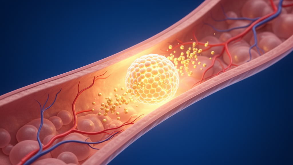
They have attracted attention as regenerative agents because they are highly biocompatible and low in toxicity, and because they can simultaneously modulate apoptosis, oxidative stress, and angiogenesis. Previous work has shown that exosomes can promote epithelial regeneration and new blood vessel growth while dampening inflammation and scar formation in wound healing, and that exosomes derived from mesenchymal stem cells exert anti-inflammatory effects and support intestinal barrier repair. Hypoxic preconditioning—growing the source cells under reduced oxygen—has been reported to make exosomes more effective in tissue repair, though direct studies of hypoxia-treated exosomes for fistula treatment have been scarce.
To test the approach, the team used 24 male Sprague-Dawley rats, each weighing about 200 grams, randomly divided into four groups of six. After a week of acclimatization, all animals underwent surgical cecostomy to establish an enterocutaneous fistula model. One group served as an untreated control, while the other three received fistula tract implantation of a gelatin sponge loaded with either lyophilized phosphate-buffered saline, hypoxia-preconditioned exosomes, or normoxic exosomes. The gelatin sponge served as a carrier because exosomes have a short half-life and are quickly cleared from the body, and gelatin is a relatively safe biomaterial suited to sustained local delivery. After 21 days of treatment, the animals were euthanized and serum and perifistulous tissue were collected for analysis.
The histological results pointed clearly toward the hypoxia-preconditioned exosome group. Hematoxylin-eosin staining showed reduced inflammatory cell infiltration in both exosome-treated groups compared with the control and buffer-sponge groups, while Masson’s trichrome staining revealed extensive blue-stained collagen fiber proliferation in the exosome-treated tissues. TUNEL staining, which flags dying cells, showed abundant positive signals in the control and buffer groups, whereas the apoptotic index in the hypoxia-exosome group was significantly lower, at a statistical threshold of P < 0.001. In other words, the treatment was associated with less inflammation, less cell death, and more of the collagen matrix that fills and closes a healing wound.
Blood tests and immune-cell analysis reinforced that picture. Enzyme-linked immunosorbent assays of serum showed that the hypoxia-exosome group had the lowest IL-1β levels and the highest IL-10 levels of all four groups. Flow cytometry of cells from the fistula tissue, using the M1 markers CD86 and iNOS and the M2 markers CD206 and ARG-1, revealed that this group had the lowest proportion of M1 macrophages—the inflammatory, microbe-clearing phenotype—and the highest proportion of M2 macrophages, the anti-inflammatory, reparative phenotype. Macrophage polarization toward the M2 state is considered crucial for tissue repair, and the authors interpret this shift as a central mechanism of the therapy’s effect.
Vascular changes were also evident. Immunofluorescence staining of perifistulous tissue showed that in the control and buffer groups, CD31-positive signals, marking endothelial cells, were abundant while α-SMA-positive signals, marking the smooth-muscle coat of mature vessels, were sparse. In the exosome-treated groups, numerous α-SMA-positive signals appeared, and most CD31 signals were closely surrounded by α-SMA, a pattern the authors read as more mature vascular structures.
In vitro experiments dissected the effects on individual repair-cell types. Human HaCaT keratinocytes treated with the gelatin sponge-loaded hypoxic exosomes showed significantly higher viability in CCK-8 assays than untreated controls, and in scratch wound assays their migration rate reached 100 percent after 24 hours, with migration significantly exceeding controls over 48 hours. Western blot analysis of human dermal fibroblasts showed significantly elevated expression of both Collagen I and Collagen III after treatment. Human dermal microvascular endothelial cells exposed to the exosomes formed denser branch connections and mesh-like structures in Matrigel tube-formation assays. Finally, RAW 264.7 macrophages treated with the exosomes showed a significantly reduced proportion of CD86-positive iNOS-positive M1 cells and a markedly increased proportion of CD206-positive ARG-1-positive M2 cells, confirmed by both flow cytometry and immunofluorescence co-localization.
The authors conclude that gelatin sponges loaded with hypoxia-preconditioned exosomes suppress inflammatory responses, enhance the function of the key cells involved in repair, promote collagen deposition and new blood vessel formation, and thereby create favorable conditions for fistula tissue repair. They frame the mechanism as multi-pronged: M2-polarized macrophages calm the local immune environment, fibroblasts deposit the collagen scaffold of granulation tissue, endothelial cells build mature vascular networks, and epithelial cells proliferate and migrate to resurface the tract. Hypoxic preconditioning, they note, has previously been reported to promote endothelial proliferation, migration, and capillary-like structure formation with superior efficacy to normoxic exosomes, and to enhance exosome yield and internalization efficiency, particularly when combined with gel-based sustained-release systems.
Important caveats remain. This was an animal study in a surgically induced rat fistula model with six animals per group, and the reported clinical trial number is listed as not applicable; no human data were generated, and the findings cannot yet be extended to patients with enterocutaneous fistulas. The study reports associations between treatment and improved histological, serological, and cellular outcomes over a 21-day window rather than long-term closure rates or safety data. The authors position the work as providing novel insights and a theoretical basis for future therapies, with the gelatin-sponge exosome platform proposed as a promising biological strategy that would require substantially more preclinical and, eventually, clinical validation before any translation to human fistula management.
Subject of Research: Hypoxia-preconditioned exosomes delivered on gelatin sponges as an experimental therapy for enterocutaneous fistulas in rats
Article Title: Hypoxia-pretreated exosomes promote enterocutaneous fistula healing via regulating inflammation and tissue repair
Article References: Liu, Y., Zhu, B., Liu, H., Yuan, Q., Yuan, C., Meng, Y., Zhou, H., Sun, L., Liu, B., & Wu, Z. (2026). Hypoxia-pretreated exosomes promote enterocutaneous fistula healing via regulating inflammation and tissue repair. BMC Molecular and Cell Biology, 27(1), Article 35. https://doi.org/10.1186/s12860-026-00592-w
Image Credits: AI Generated
DOI: 10.1186/s12860-026-00592-w
Keywords: exosomes, hypoxia preconditioning, enterocutaneous fistula, gelatin sponge, macrophage polarization, M2 macrophages, collagen deposition, angiogenesis, rat model, wound healing, IL-10, IL-1β
News Source: Drew Townsend. (October 29, 2026). Hypoxia-Preconditioned Exosomes Speed Fistula Healing in Rat Study. Scienmag.