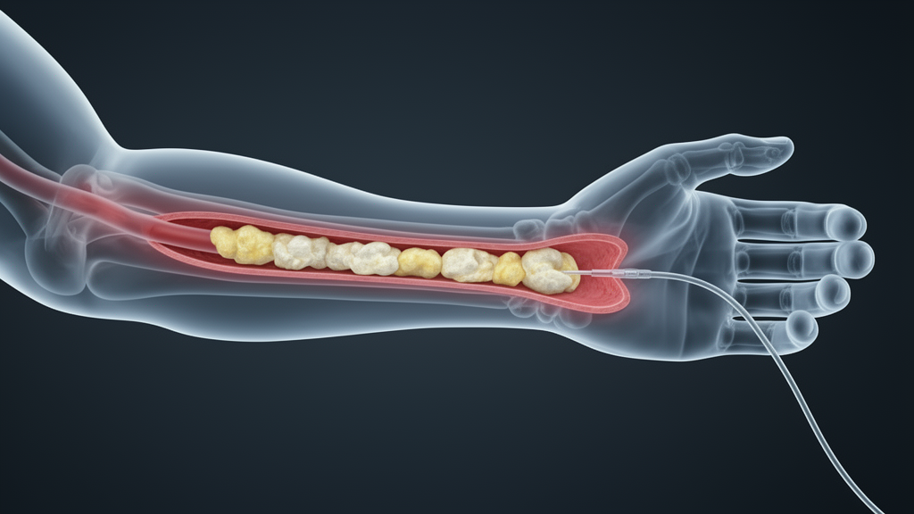
In a retrospective single-center study, researchers at Beijing Luhe Hospital, Capital Medical University, analyzed radial artery OCT images from 461 patients who underwent transradial percutaneous coronary intervention between January 2021 and August 2025. Their findings, published in Frontiers in Medicine, show that OCT-defined radial artery calcification was present in 61 of the 461 patients, or 13.2 percent of the cohort.
The study’s central observation concerns where calcification tends to occur along the radial artery. The researchers divided the vessel into proximal, middle, and distal segments and found a clear gradient: calcification involved the proximal segment in 17 patients (3.7 percent), the middle segment in 25 patients (5.4 percent), and the distal segment in 52 patients (11.3 percent).
Beyond its location, the character of the calcified deposits varied considerably. Among the 214 calcified lesions identified across the cohort, spotty calcification was the most common morphology, accounting for 151 lesions, or 70.6 percent. This heterogeneity suggests that radial artery calcification is not a single uniform entity but a spectrum of patterns that OCT can distinguish.
To understand what factors are associated with the presence of radial artery calcification, the team conducted exploratory patient-level analyses. All five radial artery plaque features they examined were associated with calcification after multivariable adjustment, and these associations remained statistically significant after correction for multiple comparisons using the Benjamini–Hochberg false discovery rate method. Notably, these associations involved noncalcified plaque features, indicating that calcification frequently coexists with other forms of radial artery atherosclerosis rather than appearing in isolation.
The investigators also looked at whether calcification in the radial artery reflects calcification elsewhere in the vascular system. In multivariable analysis, three factors remained independently associated with radial artery calcification: older age, diabetes mellitus, and the presence of a coronary calcific nodule on coronary OCT. The coronary calcium score, by contrast, was not statistically associated with radial artery calcification after adjustment. This distinction is of interest because it suggests that the presence of a specific coronary lesion type, the calcific nodule, may be a more informative correlate of radial artery calcification than a global coronary calcium burden measure.
The authors emphasize that the frequency, distribution, and OCT features of radial artery calcification had previously remained poorly defined, which is the gap this study was designed to address.
Several limitations of the study should be kept in mind when interpreting the results. It was a single-center retrospective observational study, meaning that the 461 patients were drawn from one hospital and that the analysis relied on images and data already collected during routine care. The patient-level analyses of plaque features were explicitly described as exploratory, and the findings represent associations within this cohort rather than causal effects. The cohort consisted of patients undergoing transradial PCI, so the results may not generalize to populations without coronary artery disease or to other centers with different patient demographics.
Within those constraints, the study offers a detailed quantitative picture of radial artery calcification as seen through OCT: it affects roughly one in eight patients in this cohort, clusters in the distal segment, most often takes a spotty form at the lesion level, and is associated with older age, diabetes, and coronary calcific nodules. The authors also report that exploratory analyses linked radial artery calcification to multiple noncalcified radial artery plaque features, suggesting that calcification is embedded within a broader pattern of radial artery atherosclerosis.
Subject of Research: Optical coherence tomography imaging of radial artery calcification in patients undergoing transradial coronary intervention
Article Title: Optical coherence tomography–defined radial artery calcification: frequency, phenotypes, and correlates in a retrospective transradial PCI cohort
Article References: He, S., Zhou, J., Liu, H., Wang, H., Li, Z., & Guo, J. (2026). Optical coherence tomography–defined radial artery calcification: frequency, phenotypes, and correlates in a retrospective transradial PCI cohort. Frontiers in Medicine, 13, Article 1945441. https://doi.org/10.3389/fmed.2026.1945441
Image Credits: AI Generated
DOI: 10.3389/fmed.2026.1945441
Keywords: radial artery calcification, optical coherence tomography, transradial access, percutaneous coronary intervention, vascular calcification, coronary calcific nodule, spotty calcification, diabetes mellitus, atherosclerosis, medical imaging, Optical, coherence
News Source: Ophelia Keating. (October 27, 2026). Radial artery calcification found in 13 percent of transradial PCI patients. Scienmag.