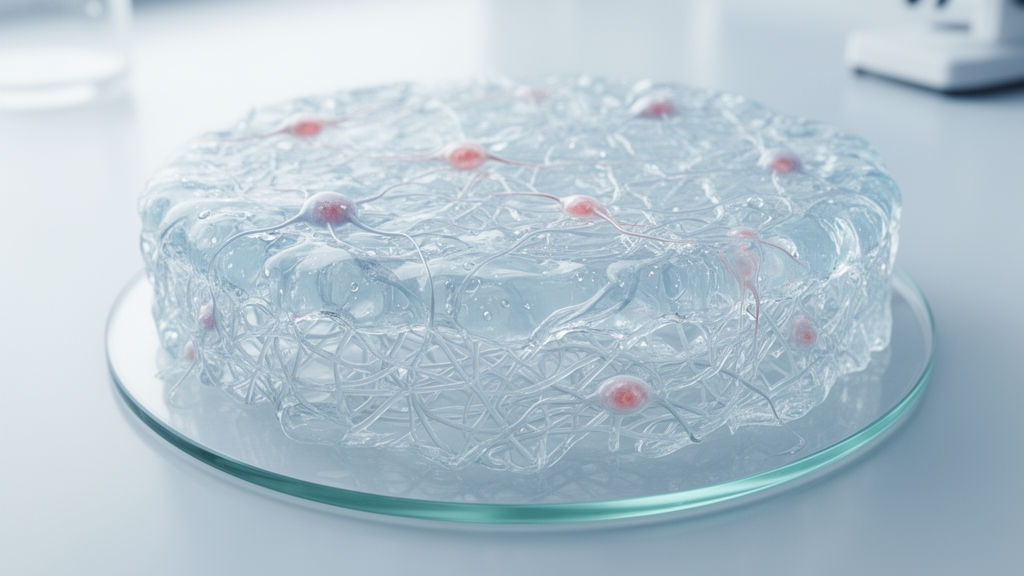
The appeal of hydrogels in regenerative medicine begins with a deceptively simple physical observation: soft tissues of the human body are largely water. Cartilage, for instance, contains roughly seventy to eighty percent water by weight, while the brain and spinal cord are even more hydrated. Any material intended to interface with, replace, or regenerate such tissues must therefore operate comfortably in an aqueous environment without provoking inflammation or mechanical mismatch. Hydrogels, three-dimensional networks of hydrophilic polymer chains that can absorb and retain large volumes of water while maintaining structural integrity, satisfy this requirement in a way that few other material classes can. Their water-rich character reduces interfacial friction with surrounding tissue, permits diffusion of nutrients and metabolic waste, and allows elastic behavior that approximates the compliance of native biological structures. This is why the extracellular matrix analogy that dominates the hydrogel literature is not merely rhetorical convenience but a genuine design principle: a well-engineered hydrogel can reproduce the hydrated, compliant, porous microenvironment in which cells naturally reside.
The distinction between physically crosslinked and chemically crosslinked hydrogels deserves closer attention, because it shapes nearly every downstream decision in scaffold design. Physically crosslinked networks rely on reversible interactions such as hydrogen bonding, ionic bridging, hydrophobic association, or host-guest complexation. Alginate, for example, gels almost instantaneously when its guluronic acid blocks coordinate divalent calcium ions, a property that has made it a mainstay of cell encapsulation and bioprinting, since cells can be mixed into the precursor solution and entrapped gently as the network forms around them. The reversibility of these bonds, however, means the gel can erode, swell excessively, or exchange ions with the surrounding physiological fluid, which limits long-term mechanical stability. Chemically crosslinked networks, formed through covalent reactions such as radical polymerization, click chemistry, or enzymatic coupling, offer superior dimensional stability and tunable degradation, but the crosslinking chemistry must be chosen with care because reactive intermediates, residual monomers, or ultraviolet initiation can damage encapsulated cells. The most successful modern platforms often combine both modes, using rapid physical gelation to shape the material in situ and slower covalent crosslinking to lock in final properties.
Among natural polymers, each candidate brings a distinct biological personality. Collagen, the most abundant protein in the mammalian body, provides inherent cell-binding motifs such as the RGD sequence that synthetic materials must otherwise graft on artificially. Gelatin, its partially denatured derivative, retains many of these motifs while being easier to process and chemically modify, and its thermoreversible gelation near physiological temperatures underpins its use in injectable carriers and bioinks. Hyaluronic acid, a linear glycosaminoglycan, is not merely a passive structural component but an active signaling molecule: it interacts with cell surface receptors such as CD44 and RHAMM, influencing migration, proliferation, and inflammation resolution, which explains its prominence in wound healing and neural applications. Chitosan, derived from chitin, carries positive charges under physiological conditions, enabling electrostatic complexation with DNA, RNA, and negatively charged growth factors, a feature exploited in gene delivery and antimicrobial formulations. Alginate, by contrast, is relatively bioinert toward mammalian cells, which is a double-edged characteristic: it minimizes nonspecific immune activation but must be functionalized if specific cell adhesion is desired.
Synthetic polymers answer a different set of needs. Polyethylene glycol remains the workhorse of synthetic hydrogel chemistry because it is highly hydrated, essentially protein-resistant in its unmodified form, and amenable to an extraordinary range of end-group functionalizations. This inertness allows researchers to build matrices from a blank slate, adding only the biochemical cues they deliberately choose, which is invaluable for systematically studying how matrix composition affects cell behavior. Polycaprolactone, with its slow degradation profile spanning months to years, suits applications requiring prolonged mechanical support, such as bone regeneration, while polylactic acid and its copolymers degrade faster and can be tuned through stereochemistry and molecular weight. Poly-N-isopropylacrylamide exhibits a lower critical solution temperature near thirty-two degrees Celsius, meaning it transitions from a hydrated coil to a collapsed globule as temperature rises through this threshold. Formulated appropriately, such thermoresponsive polymers remain soluble when handled cold and gel in place upon reaching body temperature, an elegant solution to the injectable delivery problem that avoids cytotoxic crosslinkers entirely.
The concept of mechanical tunability merits quantitative appreciation. Cells are remarkably sensitive mechanosensors: substrate stiffness in the kilopascal range can direct mesenchymal stem cells toward neurogenic, myogenic, or osteogenic lineages even in the absence of soluble differentiation factors, a finding that reshaped how the field thinks about stem cell culture. Brain tissue is on the order of a few hundred pascals in stiffness, muscle roughly ten to twenty kilopascals, and mineralized bone several gigapascals. A hydrogel intended to nurture neural progenitors must therefore be dramatically softer than one designed to support osteoblast maturation, and the ability to dial in elastic modulus through polymer concentration and crosslink density is among the most powerful levers in hydrogel engineering. Stress relaxation, the rate at which a matrix dissipates applied strain, has emerged as an equally important parameter, because dynamically remodeling matrices permit cells to spread, migrate, and deposit their own extracellular matrix in ways that purely elastic networks of identical stiffness do not.
Injectable hydrogel systems address one of the most practical constraints in regenerative surgery: the difficulty of pre-formed implants to conform to irregular defect geometries. A preformed scaffold requires invasive implantation, risks poor contact with surrounding tissue, and cannot easily fill complex cavities. In situ gelling systems, delivered as liquid precursors through minimally invasive injection, conform to the defect shape and polymerize under physiological triggers such as temperature, pH, ion concentration, or light exposure. Shear-thinning formulations add another layer of sophistication: they flow readily under the high shear of the injection needle yet recover their mechanical integrity immediately upon deposition, protecting encapsulated cells from the transient stresses of delivery. This property, borrowed from biological gels such as mucus and the cytoskeleton, has proven particularly valuable for bioprinting applications where the printed filament must hold its shape while remaining processable.
Nanocomposite hydrogels illustrate how inorganic phases can endow soft networks with capabilities they otherwise lack. Incorporating nanoscale hydroxyapatite into polymer matrices introduces mineral surfaces that promote osteoblast adhesion and osteogenic differentiation while simultaneously stiffening the network toward bone-like mechanics. Graphene oxide and carbon nanotubes confer electrical conductivity, which matters enormously for cardiac and neural engineering, since these tissues rely on synchronized electrical signaling that insulating hydrogels cannot support. Silica and clay nanoparticles can serve as physical crosslinking hubs, dramatically increasing toughness through sacrificial bond mechanisms in which nanoparticle-polymer interactions break and reform under load, dissipating energy that would otherwise fracture the network. The design challenge lies in ensuring that the reinforcing phase remains biocompatible, does not leach, and does not aggregate, since nanoparticle clustering creates stress concentrations that undermine the very toughness the fillers are meant to provide.
Stimuli-responsive behavior extends beyond temperature. pH-responsive systems exploit the acidic microenvironments of tumors, inflamed tissue, or infected wounds to trigger drug release precisely where it is needed. Redox-responsive networks disassemble in the reducing cytosol, enabling intracellular delivery of nucleic acids and proteins. Photo-responsive formulations allow spatiotemporally precise actuation, since light can be focused, patterned, and switched with exceptional control, and visible or near-infrared wavelengths penetrate tissue with minimal phototoxicity. Enzyme-responsive hydrogels represent perhaps the most biologically elegant category: by incorporating peptide sequences cleaved by matrix metalloproteinases, the scaffold degrades only where and when cells actively remodel it, mimicking the natural balance between matrix deposition and proteolytic turnover that governs tissue morphogenesis.
Self-healing hydrogels tackle a chronic failure mode of soft biomaterials: crack propagation under cyclic loading. Native tissues remodel continuously and repair microdamage; conventional synthetic gels accumulate defects until they fail catastrophically. Networks engineered with reversible crosslinks, dynamic covalent bonds such as boronate esters and imine linkages, or supramolecular host-guest interactions can autonomously re-form broken connections, restoring function after damage. In load-bearing cartilage applications, where joints experience millions of loading cycles annually, this resilience is not a luxury but a prerequisite for clinical durability.
The convergence of hydrogels with drug and gene delivery has produced some of the field’s most clinically near-term applications. Because hydrogels can entrap therapeutic molecules within their hydrated mesh and release them through diffusion, swelling, or degradation kinetics, they function as depot formulations that maintain therapeutic concentrations over days to months while avoiding the peaks and troughs of systemic administration. Growth factor delivery illustrates the principle: bone morphogenetic proteins, vascular endothelial growth factor, and transforming growth factor-beta each orchestrate distinct phases of tissue repair, and hydrogel carriers can be engineered to release them sequentially, mirroring the temporal choreography of natural healing. Affinity-based release, in which growth factors bind reversibly to polymer-bound heparin or other binding motifs, smooths release profiles far better than simple diffusion. Gene delivery from hydrogels, using plasmid DNA, messenger RNA, or viral vectors complexed with cationic polymers, aims to recruit resident cells into local production of therapeutic proteins, converting the scaffold from a passive carrier into an active instruction set.
Biosensing represents a frontier where hydrogels act as both transducer and interface. Their water content and softness minimize fouling and immune reaction at sensor-tissue interfaces, historically the limiting factor in continuous glucose monitoring and other implantable sensors. Conductive hydrogels incorporating PEDOT, polyaniline, or metallic nanostructures enable low-impedance recording and stimulation of electrogenic tissue, while responsive swelling or degradation can transduce biochemical signals into optical or electrical readouts. The same material that regenerates tissue can thus report on its progress, closing the loop for adaptive therapy.
The translational barriers documented in the recent literature deserve sober acknowledgment. Preclinical animal models frequently fail to predict human outcomes, a problem the FDA Modernization Act 2.0 partially addresses by permitting alternatives to animal testing for certain regulatory purposes, opening space for organoid platforms and advanced in vitro systems built on hydrogel matrices that better recapitulate human physiology. Scale-up introduces its own difficulties: laboratory syntheses relying on precise stoichiometry, sterile handling, and gentle crosslinking conditions do not always translate to good manufacturing practice environments. Sterilization poses a particular dilemma, since autoclaving, gamma irradiation, and ethylene oxide can each alter polymer molecular weight, crosslink density, or bioactivity. Regulatory classification adds further complexity, because combination products that merge device, drug, and biologic elements face overlapping review pathways. Progress will likely require not only better materials but better standardized characterization, reproducible manufacturing, and harmonized preclinical evaluation frameworks, so that the remarkable laboratory performance of next-generation hydrogels can be converted into dependable clinical benefit.
Subject of Research: Advanced hydrogels for tissue engineering and regenerative medicine: from material design to clinical translation
Article Title: Advanced hydrogels for tissue engineering and regenerative medicine: from material design to clinical translation
Article References: Vyas, J., Raytthatha, N., Vyas, P., Shah, N., & Modi, D. (2026). Advanced hydrogels for tissue engineering and regenerative medicine: from material design to clinical translation. Frontiers in Biomaterials Science, 5, Article 1861958. https://doi.org/10.3389/fbiom.2026.1861958
Image Credits: AI Generated
DOI: 10.3389/fbiom.2026.1861958
Keywords: hydrogels, tissue engineering, regenerative medicine, biomaterials, extracellular matrix, injectable hydrogels, stem cells, 3D bioprinting, drug delivery, clinical translation, Advanced, tissue
News Source: Matthew Wilson. (October 17, 2026). Hydrogels That Heal: How ECM-Mimicking Biomaterials Are Rewriting Regenerative Medicine. Scienmag.