Melanoma remains one of the most formidable cancers in medicine. Although it accounts for only about one percent of all skin cancer cases, its extraordinary capacity for metastasis and its stubborn resistance to conventional chemotherapy make it disproportionately deadly. Researchers have long relied on flat, two-dimensional cell cultures and animal models to test new drugs, but these systems fail to capture the dense, layered, and chemically complex environment in which real tumors live. A new perspective article published in Frontiers in Cell Death by Lissinda H. du Plessis and colleagues at North-West University in South Africa, together with Daniel Nieto of the University of A Coruña in Spain, argues that while three-dimensional bioprinted melanoma models are rapidly maturing as a platform, the analytical methods used to read out drug effects have not kept pace, and this mismatch now threatens to undermine the very promise of the technology.
The case for better models begins with melanoma biology itself. Melanoma arises from pigment-producing melanocytes, and its development is typically initiated by ultraviolet radiation-induced DNA damage that, when repaired incorrectly, produces oncogenic mutations in key regulatory genes. Activating mutations in the BRAF and NRAS proto-oncogenes drive constitutive activation of the MAPK signaling pathway, which governs cell proliferation, differentiation, and survival. Simultaneously, loss of function in tumor suppressor genes such as CDKN2A, PTEN, and TP53 disrupts cell-cycle control, apoptosis, and genomic stability. Beyond genetics, the tumor microenvironment, populated by stromal cells, tumor-infiltrating immune cells, and tumor-associated blood vessels, plays decisive roles in tumor growth, immune evasion, and metastatic spread. Any model that hopes to predict how a patient will respond to therapy must somehow reproduce this intricate biological landscape.
Current treatments illustrate why predictive models matter so much. Surgical resection remains the cornerstone for early-stage disease, while advanced melanoma is treated with immune checkpoint inhibitors and targeted therapies. Anti-CTLA-4 antibodies block inhibitory signaling between T-cells and antigen-presenting cells, unleashing T-cell activation, cytokine release, and tumor infiltration. Anti-PD-1 and PD-L1 agents prevent the co-inhibitory ligand-receptor interaction that otherwise dampens T-cell responses. In BRAF-mutant tumors, small-molecule inhibitors such as vemurafenib and dabrafenib are combined with downstream MEK inhibitors like trametinib and cobimetinib to suppress MAPK signaling, reducing proliferation and inducing apoptosis. Yet despite these advances, intrinsic and acquired resistance to both targeted and immune-based therapies keeps melanoma a pressing public health problem, and the drugs that succeed in flat cultures or mice frequently fail in patients.
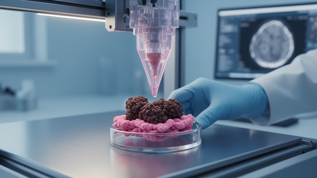
Three-dimensional bioprinting has emerged as one of the most promising strategies to close this translation gap. In this approach, a bioink, a mixture of living cells suspended in biocompatible hydrogel biomaterials, is deposited layer by layer according to a pre-determined computer-aided design, building melanoma constructs that more closely resemble skin tissue architecture and better capture clinical heterogeneity. The hydrogels provide structural support, protect cells during printing, and allow the incorporation of multiple cell types, including melanoma cells, fibroblasts, keratinocytes, endothelial cells, and immune cells, to mimic the tumor microenvironment. Several printing technologies are in use, including laser-assisted, inkjet-based, stereolithography-based, and extrusion-based bioprinting. Pneumatic extrusion bioprinting has become the most widely adopted because of its accessibility, cost-effectiveness, and capacity to replicate tissue complexity, while digital light processing offers the added benefit of fabricating complex structures with high resolution. Importantly, the technology also aligns with the principles of replacement, reduction, and refinement in animal research and opens the door to personalized, patient-specific tumor models.
The field has already produced impressive demonstrations. Organotypic melanoma spheroid skin models built from primary keratinocytes and fibroblasts combined with metastatic melanoma spheroids have shown epidermal differentiation, basal lamina formation, and necrotic cores that mimic human disease. High-throughput drug screening on NRAS-mutant and multicomponent melanoma spheroids has identified candidate inhibitors such as daunorubicin hydrochloride and pyrvinium pamoate. Patient-derived organoids from melanoma brain metastases, cutaneous tumors, and mucosal melanoma have preserved key histological features and mutational profiles and responded to agents including dabrafenib plus trametinib and temozolomide. Microfluidic melanoma-on-a-chip systems have distinguished carboplatin-responsive from carboplatin-resistant tumors, and bioprinted constructs have recreated vascular-lymphatic interactions to study metastasis. More recently, bioprinted A375 melanoma constructs have been validated for apoptotic cell death analysis using flow cytometry, showing compatibility with quantitative mechanistic assays used in preclinical research.
Yet the du Plessis team’s central argument is that the bottleneck has shifted from building the models to measuring what happens inside them. Most biochemical and molecular techniques used to assess cell viability, proliferation, drug efficacy, and toxicity, including luminescence, colorimetric, and fluorometric assays, microscopy, flow cytometry, quantitative polymerase chain reaction, and SDS-PAGE, were optimized for cells growing in flat monolayers. Applied to dense, multilayered 3D matrices, they often yield inconsistent or misleading results because of matrix interference, diffusion limitations, and uneven cell distribution. Bioprinted constructs frequently exceed 500 micrometers in thickness, and diffusion barriers restrict reagent penetration to deep layers, which can cause an underestimation of viable cells in the interior and lead to incorrect drug potency predictions and misinterpretation of pharmacodynamic behavior. Variability in construct size, shape, and cell number further complicates comparisons and can skew potency estimates between studies.
The printing process itself adds another layer of complexity. Pneumatic extrusion bioprinting damages cells through mechanical shear forces, whereas digital light processing inflicts radiative stress from light exposure and chemical stress from photoinitiators. Bioink compatibility with melanoma cells is not guaranteed either, because both bioink composition and printing conditions can alter proliferation, spheroid morphology, migration, and cell-matrix interactions even when viability is maintained. Studies of printable hydrogels such as alginate/hyaluronic acid/gelatin and pectin-nanocellulose composites have improved rheology, printability, and short-term viability of A375 melanoma cells, but these outcomes are formulation- and bioprinter-specific, meaning the same bioink may need re-optimization when transferred to different printers, extrusion parameters, or crosslinking methods. The authors call for systematic validation of identical formulations across multiple bioprinting platforms to establish true reproducibility.
Apoptosis, the regulated form of cell death that most anti-cancer drugs aim to trigger, presents its own analytical puzzles. The intrinsic, or mitochondrial, pathway is controlled by pro- and anti-apoptotic Bcl-2 family proteins that increase mitochondrial outer membrane permeability and release factors such as cytochrome c, which activates caspase-9. The extrinsic pathway is death-receptor mediated, with Fas ligand and tumor necrosis factor-alpha binding their receptors and activating caspase-8, both routes converging on effector caspase-3. Melanoma cells evade apoptosis through dysregulated Bcl-2 family proteins, overexpression of inhibitor of apoptosis proteins, and rewired pro-survival signaling through heat shock proteins. Accurately quantifying drug-induced apoptosis therefore requires comparable cell numbers or construct sizes, and the context-dependence of apoptotic signaling, shaped by cell-cell interactions, cell-matrix adhesion, biomechanical cues, and gradients of oxygen, nutrients, and drugs, is precisely what 2D cultures fail to represent. Notably, the effect of different bioprinting technologies on apoptotic signaling in melanoma remains underexplored, with current evidence limited mainly to extrusion-based systems.
Each analytical workaround carries trade-offs. Non-destructive colorimetric and fluorescence assays such as MTT, MTS, CellTiter-Glo, and resazurin-based methods preserve construct integrity but tend to return only an average response across a cell population, losing crucial biological information, and dye diffusion through the hydrogel matrix can dilute signals and distort viability readings. Destructive methods like flow cytometry require dissolving the construct into a single-cell suspension but offer rapid, quantitative, multi-parametric analysis of individual cells within heterogeneous populations, making them attractive for high-throughput drug screening. Fluorescence and confocal microscopy suffer from poor imaging depth because refractive index differences in heterogeneous hydrogels scatter light, a problem addressed through physical sectioning, tissue clearing, and refractive-index-matched hydrogels. Immunolabelling can be unreliable in thick matrices where antibodies diffuse poorly, producing weak or false-negative staining in deeper layers. Histology remains a gold standard for validating tissue structure and diagnostic markers, while lysate-based assays such as Western blotting and RT-qPCR can be biased by inefficient lysis of cells embedded in dense, highly crosslinked matrices.
The authors conclude that 3D bioprinted melanoma models genuinely represent physiologically relevant platforms capable of mimicking architectural complexity, cellular heterogeneity, and clinically relevant drug responses, but that the assays used to evaluate drug efficacy and toxicity must be re-optimized, standardized, and validated for hydrogel-embedded constructs. As high-throughput screening generates ever more parameters, the field will also need sophisticated data storage, automated analysis, miniaturization, and increasingly artificial intelligence and machine learning, all of which remain constrained by the absence of regulatory and global standards. Solving this analysis bottleneck, the researchers argue, will determine whether bioprinted melanoma models fulfill their potential to accelerate drug discovery, improve toxicity testing, and ultimately deliver safer, more effective treatments to patients battling one of medicine’s most aggressive cancers.
Subject of Research: Analytical challenges in drug efficacy and toxicity screening using 3D bioprinted melanoma models
Article Title: Challenges in drug efficacy and toxicity screening with 3D bioprinted melanoma models
Article References: du Plessis, L. H., Myeza, N., Gouws, C., Kotzé, A. F., & Nieto, D. (2026). Challenges in drug efficacy and toxicity screening with 3D bioprinted melanoma models. Frontiers in Cell Death, 5, Article 1821035. https://doi.org/10.3389/fceld.2026.1821035
Image Credits: AI Generated
DOI: 10.3389/fceld.2026.1821035
Keywords: 3D bioprinting, melanoma, drug screening, apoptosis, tumor microenvironment, bioinks, hydrogels, flow cytometry, cell viability assays, skin cancer, drug resistance, preclinical models
News Source: Nathaniel Bowman. (October 16, 2026). 3D-Printed Melanoma Models Promise Better Drug Tests but Face an Analysis Bottleneck. Scienmag.