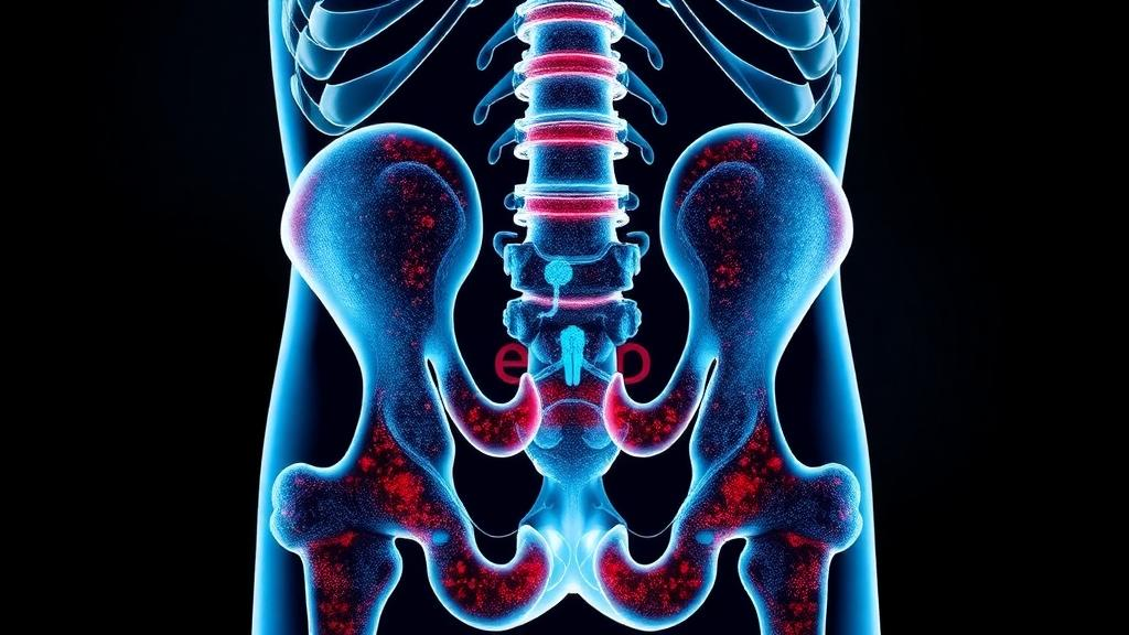
Postmenopausal osteoporosis affects an estimated 200 million women worldwide, and one in three women over the age of fifty will eventually suffer an osteoporotic fracture. For decades, treatment has centered on hormone replacement or bisphosphonates, both of which carry long-term risks. Now, a team of researchers has reported a strikingly different approach: restoring a single molecule that the body already produces. In a study published in the Journal of Advanced Research, investigators led by Chenying Zeng and Ziji Zhang identified hyodeoxycholic acid, a secondary bile acid manufactured by gut bacteria, as a key metabolic factor that declines sharply in postmenopausal osteoporosis and, when replenished, appears to rejuvenate the stem cells that keep bone strong.
The story begins in the bone marrow. Bone marrow mesenchymal stromal cells, or BMSCs, are the functional workhorses of the skeletal microenvironment, capable of becoming bone-forming osteoblasts or fat-storing adipocytes. When estrogen levels collapse after menopause, these cells undergo a pathological identity crisis: they senesce, arresting their cell cycles and secreting inflammatory factors, while simultaneously drifting toward abnormal fat production. The result is a marrow increasingly filled with fat instead of bone, a hallmark of the disease. The researchers asked whether circulating bile acids, long known as digestive detergents but increasingly recognized as metabolic signaling molecules, might govern this fate decision.
To find out, the team profiled serum bile acids in healthy donors and postmenopausal osteoporosis patients using ultra-high-performance liquid chromatography coupled with tandem mass spectrometry. The comparison revealed a distinct metabolic signature: several bile acids were altered, but hyodeoxycholic acid showed the largest and most consistent reduction in patients. When the researchers stratified patients by their serum levels, those in the low-HDCA group had BMSCs that stained far more strongly for senescence-associated beta-galactosidase and accumulated more lipid droplets during adipogenic induction than cells from high-HDCA patients. Spearman correlation analysis confirmed negative associations between HDCA levels and both senescence and fat accumulation, with tertile stratification showing a dose-dependent relationship.
The next question was whether the molecule could actively reverse the damage, not merely mark it. The researchers established an inflammatory senescence model by exposing patient-derived BMSCs to hydrogen peroxide, then treating them with HDCA at 10, 20, and 40 micromolar concentrations. The results were unambiguous: HDCA dose-dependently reduced senescence staining, suppressed the senescence effectors p53, p21, and p16, and lowered the adipogenic marker FABP4. EdU incorporation assays showed that the compound also rescued the proliferative capacity that oxidative stress had destroyed. Notably, HDCA produced the same protective effects in naturally aged, late-passage BMSCs, suggesting the benefit extends beyond a single laboratory model of injury.
Mechanistically, the team traced a signaling chain that runs from the cell membrane to the nucleus and finally to the mitochondria. Molecular docking simulations predicted that HDCA binds the membrane bile acid receptor TGR5 with higher affinity than the nuclear receptor FXR, and microscale thermophoresis confirmed preferential binding, with a dissociation constant of 26.5 micromolar for TGR5 versus 61.7 micromolar for FXR. As a G protein-coupled receptor, TGR5 activation stimulates cyclic AMP production and protein kinase A signaling. In the senescent BMSCs, HDCA restored TGR5 expression, elevated intracellular cAMP, and increased phosphorylated PKA. Pharmacological blockade with the PKA inhibitor H89 abolished HDCA’s protection, while the TGR5 agonist INT-777 enhanced it, and siRNA knockdown of TGR5 similarly blunted the effect.
Downstream of PKA, an unbiased network pharmacology analysis pointed to a surprising nuclear partner: ESR1, the estrogen receptor alpha. HDCA dose-dependently reversed the hydrogen-peroxide-driven loss of ESR1 protein, but this upregulation failed when PKA was inhibited, placing ESR1 firmly downstream of the TGR5-cAMP-PKA axis. Immunofluorescence showed increased nuclear accumulation of ESR1 after HDCA treatment. When the researchers silenced ESR1 with siRNA, HDCA could no longer suppress lipid accumulation or senescence markers, and p21 rose sharply. In essence, HDCA appears to recapitulate part of estrogen’s protective program in bone stem cells through a non-hormonal route, a finding with obvious implications for patients who cannot take hormone therapy.
The final piece of the mechanism lies in mitochondrial quality control. RNA sequencing of HDCA-treated cells revealed downregulation of p53 pathway genes and enrichment of terms related to oxidative phosphorylation and mitochondrial electron transport. Functional assays showed that HDCA increased colocalization between the autophagosome marker GFP-LC3 and mitochondria labeled with mito-RFP, indicating active mitophagy, the selective disposal of damaged mitochondria. HDCA upregulated the mitophagy mediators PINK1 and Parkin and promoted p62 degradation. A time-course analysis revealed that LC3-II conversion occurred within 12 to 24 hours, while recovery of mitochondrial membrane potential followed at 24 to 48 hours, suggesting that clearing damaged mitochondria precedes functional restoration. Blocking mitophagy with Mdivi-1 abolished HDCA’s protective effects, confirming the pathway’s necessity.
To test whether these cellular findings translate to a living organism, the team used an ovariectomized mouse model that mimics postmenopausal bone loss. After eight weeks of oral HDCA supplementation at 80 milligrams per kilogram per day, micro-CT imaging showed substantially improved trabecular bone volume fraction, bone mineral density, and trabecular number, effects comparable to the clinical bisphosphonate alendronate used as a positive control. Histology revealed that the massive marrow fat infiltration typical of estrogen-deficient mice was markedly suppressed, and immunohistochemistry confirmed reduced p53 and FABP4 signaling in femoral tissue. HDCA also restored TGR5 and ESR1 expression in both intestinal and bone tissues, lowered serum IL-6, and reversed the serum HDCA deficit, implicating the gut-bone axis in the systemic response. Preliminary safety assessments showed no significant changes in body weight, organ indices, or liver and kidney markers over the eight-week treatment.
The authors are careful to frame the work as a foundation rather than a finished therapy. They note that the upstream link between estrogen deficiency and reduced intestinal HDCA production remains untraced, that the in vivo data cannot yet distinguish effects on BMSCs from direct actions on osteoblasts and osteoclasts, and that the mitophagy inhibitor used lacks full specificity, so genetic validation targeting PINK1 or Parkin is still needed. Pharmacokinetics and long-term safety, including effects on the bile acid pool and gut microbial ecology, await dedicated study. The human equivalent dose estimated by allometric scaling, roughly 390 milligrams per day for a 60-kilogram adult, is a theoretical projection that requires formal clinical testing.
Even with those caveats, the study represents a compelling convergence of metabolomics, stem cell biology, and translational medicine. It positions a gut microbiota-derived metabolite as an endogenous regulator of stem cell fate, unifies senescence and aberrant adipogenesis as two faces of the same mitochondrial defect, and demonstrates that a single oral supplement can restore both signaling and structure in a disease model. If future clinical trials validate these findings, the millions of women facing postmenopausal bone loss may one day have an option that works not by forcing bone to grow or blocking its breakdown, but by coaxing their own aging stem cells back to youthfulness from within.
Subject of Research: Bile acid metabolism and bone marrow mesenchymal stem cell rejuvenation in postmenopausal osteoporosis
Article Title: Hyodeoxycholic acid rejuvenates BMSCs against postmenopausal osteoporosis by attenuating senescence and aberrant adipogenesis via TGR5-ESR1-mediated mitophagy
Article References: Zeng, C., Yu, W., Kang, Y., Liu, W., Chen, Z., Xiao, Z., Liu, Z., Zhang, W., Li, Q., Zeng, Y., Li, Z., Pan, B., Xie, Z., & Zhang, Z. (2026). Hyodeoxycholic acid rejuvenates BMSCs against postmenopausal osteoporosis by attenuating senescence and aberrant adipogenesis via TGR5-ESR1-mediated mitophagy. Journal of Advanced Research. https://doi.org/10.1016/j.jare.2026.10.020
Image Credits: AI Generated
DOI: 10.1016/j.jare.2026.10.020
Keywords: postmenopausal osteoporosis, hyodeoxycholic acid, bile acids, bone marrow mesenchymal stem cells, TGR5, ESR1, mitophagy, PINK1, Parkin, gut-bone axis, cellular senescence, adipogenesis
News Source: Beatrice Stafford. (October 11, 2026). Gut Bile Acid Rejuvenates Aging Bone Stem Cells to Fight Postmenopausal Osteoporosis. Scienmag.