Chronic liver disease remains one of the most formidable challenges in modern medicine, claiming roughly two million lives worldwide each year and progressing silently toward cirrhosis and hepatocellular carcinoma in millions more. Whether triggered by autoimmune attack, alcohol, metabolic dysfunction or viral hepatitis, the end point is often the same: progressive scarring that irreversibly compromises liver function. Yet effective therapeutic options remain stubbornly limited. A comprehensive review published in Experimental & Molecular Medicine by Yinuo Liu, Baichao Yu, Qiaoyan Liu and colleagues at Renji Hospital, Shanghai Jiao Tong University School of Medicine, now argues that a decisive piece of the puzzle lies far from the liver itself, in the trillions of microbes inhabiting the intestine and their intricate dialogue with T lymphocytes, the central effectors of adaptive immunity.
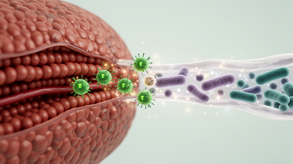
The anatomical logic behind this connection is elegant. The liver receives the entire venous outflow of the gut through the portal circulation, meaning it is continuously bathed in microbial products, metabolites and dietary antigens. In return, the liver secretes bile acids and other bioactive mediators into the intestine that directly shape which microbial species can thrive there. This bidirectional communication network, known as the gut–liver axis, places the immune system at its centre, tasked with maintaining a delicate equilibrium between tolerance toward commensal bacteria and robust defence against pathogens. T cells are uniquely positioned within this axis because they can recognize an extraordinarily broad spectrum of microbial antigens, and their differentiation, function and plasticity are in turn profoundly sculpted by microbial cues.
The review details how specific gut microbes direct the fate of distinct T helper subsets with remarkable precision. Segmented filamentous bacteria, for example, are a classical inducer of Th17 cells: their adhesion to intestinal epithelial cells triggers secretion of serum amyloid A, which acts on CD11c-positive cells to potentiate Th17 differentiation, amplified by a positive feedback loop involving interleukin-1β. More recently, the ubiquitous commensal Alcaligenes faecalis was shown to induce intestinal Th17 cells through the Trim21–Fbxw7 axis, and even the fungus Candida albicans can promote Th17 accumulation in the colon. Bacterial metabolites add a second layer of control. Short-chain fatty acids such as acetate, propionate and butyrate, secondary bile acids including deoxycholic acid and isolithocholic acid, and tryptophan-derived indoles all modulate Th17, Th1 and regulatory T cell programmes, often through inhibition of histone deacetylases or direct engagement of transcription factors such as RORγt.
Regulatory T cells, which express the transcription factor FOXP3 and enforce immune tolerance, are equally dependent on microbial instruction. Genera including Clostridium, Bacteroides, Bifidobacterium and Helicobacter promote colonic Treg induction, while Akkermansia muciniphila alleviates colonic inflammation by expanding RORγt-positive Treg responses. Butyrate and propionate enhance Foxp3 expression through histone acetylation at conserved non-coding sequences, and specific bile acid metabolites such as 3β-hydroxydeoxycholic acid promote Treg generation. Beyond conventional T cells, the review highlights innate-like lymphocytes with deep microbial entanglement: mucosal-associated invariant T cells depend on early-life exposure to riboflavin-synthesizing commensals, which produce the potent antigen 5-OP-RU, while hepatic natural killer T cells are regulated by microbiota-driven bile acid metabolism that controls CXCL16 expression in liver sinusoidal endothelial cells. Gamma-delta T cells, abundant in both liver and intestine, respond to microbial lipid antigens presented by hepatocytes and modulate their interleukin-17 output according to bacterial load.
Crucially, the communication is not one-way. T cells themselves reshape the microbial ecosystem. Microbiota-derived extracellular ATP constrains T follicular helper cell function through the P2X7 receptor, limiting secretory IgA production; in P2X7-deficient mice, excessive IgA disrupts microbial composition and drives metabolic dysregulation. Gamma-delta T cell-deficient mice display altered microbiota composition that feeds back into immune dysregulation, and studies of NKT cell-deficient animals have reported shifts in commensal communities, although recent work using littermate controls suggests maternal transmission and caging conditions may matter more than genotype. T cell plasticity further complicates the picture: Th17 cells can convert toward Th1 or T follicular helper phenotypes, and while the initial conversion of CD8-positive to CD4-positive T cells can occur without microbes, their subsequent homing and expansion in the gut critically depend on microbial signals.
When this finely balanced system fails, the consequences for the liver can be severe. The review describes how disruption of the intestinal barrier, the so-called leaky gut, permits bacteria, toxins and microbial antigens to translocate into the portal vein and systemic circulation. Molecular mimicry provides one mechanistic bridge: the pyruvate dehydrogenase complex E2 of Escherichia coli cross-reacts with its human counterpart at the T cell receptor level, contributing to primary biliary cholangitis, while cross-reactivity between microbial antigens and host components has been implicated in primary sclerosing cholangitis and autoimmune hepatitis. Gut dysbiosis, characterized by reduced microbial diversity, overgrowth of pathogenic taxa and depletion of beneficial commensals, accompanies virtually every form of chronic liver disease, and advanced cirrhosis additionally enriches virulence factors and antimicrobial resistance genes within the microbiome.
Disease-specific examples illustrate the diversity of these pathogenic axes. In autoimmune hepatitis, the pathobiont Enterococcus gallinarum translocates to the liver, where its DNA has been detected in patients and where it drives Th17 responses and autoantibody production in mice; conversely, the microbial metabolite indole-3-carboxaldehyde ameliorates immune-mediated hepatitis by activating the aryl hydrocarbon receptor in T cells. In primary sclerosing cholangitis, Klebsiella pneumoniae with strain-specific epithelial pore-forming capacity breaches the intestinal barrier and provokes robust hepatic Th17 responses, while translocated Lactobacillus gasseri induces IL-17A production by gamma-delta T cells in cholestatic models. In alcohol-associated liver disease, ethanol promotes fungal dysbiosis and generates Candida albicans-specific Th17 cells that migrate to the liver and exacerbate injury through IL-17 signalling on Kupffer cells, while peripheral MAIT cells become depleted and hyperactivated through heightened exposure to translocated bacterial products.
Metabolic dysfunction-associated steatotic liver disease, which affects approximately 38 percent of the global adult population, adds yet another dimension. High-fat diet models show depletion of protective intestinal Th17 cells, promoting microbial migration that accelerates disease, while microbiota-derived lipid antigens presented by hepatocytes via CD1d stimulate hepatic IL-17-producing gamma-delta T cells. Faecal bacterial extracts from patients with MASLD-related hepatocellular carcinoma induce immunosuppressive phenotypes ex vivo, expanding Treg cells and attenuating CD8-positive T cell function in a short-chain fatty acid-dependent manner. In chronic hepatitis B, bacterial extracts from non-cirrhotic patients promote Th17 expansion whereas those from cirrhotic patients inhibit Th1 cells, and strikingly, a probiotic consortium of Bifidobacterium longum, Enterococcus faecalis and Lactobacillus acidophilus, together with its metabolite spermidine, suppresses HBV replication by augmenting IFNγ-positive CD4-positive T cell immunity.
These mechanistic insights are now feeding into a therapeutic pipeline that targets both sides of the axis. Microbiota-directed interventions include probiotics such as Lactobacillus acidophilus, which attenuated cholestatic liver damage in mice and improved liver function in cholestatic patients, and Lactobacillus rhamnosus GG, which alleviated liver injury in moderate alcohol-associated hepatitis. Prebiotics such as inulin and camu-camu have shown benefits in metabolic liver disease, although inulin failed to improve early alcoholic liver injury despite reshaping the microbiome. Faecal microbiota transplantation increased bacterial diversity and reduced alkaline phosphatase in primary sclerosing cholangitis and restored intestinal permeability in MASLD, though it did not improve hepatic fat fraction or insulin resistance. On the T cell side, chimeric antigen receptor T cell therapy eliminated pathogenic PD-1-positive CD8-positive T cells that drive hepatocyte pyroptosis in autoimmune liver disease models, autologous Treg infusion significantly reduced liver enzymes in a patient with refractory PSC/UC, and low-dose interleukin-2 therapy that selectively expands Tregs has shown early promise.
The authors caution that the gut–liver–immune network is dynamic and bidirectional, meaning that targeting a single component may trigger unanticipated cascades; indeed, the efficacy of CAR-T therapy itself can be modulated by the intestinal microbiota. They argue that future progress will require patient-stratified approaches, deeper elucidation of the specific microbial taxa and immunological mechanisms underlying each disease, and clinical validation of candidate targets. If that integration of microbiome science with translational immunology succeeds, the microbial cues that currently fuel chronic liver disease could be repurposed as precise levers for its prevention and treatment, opening a genuinely new therapeutic frontier for conditions that have long defied conventional intervention.
Subject of Research: Bidirectional interactions between gut microbiota and T cell immunity in the progression of chronic liver disease
Article Title: Microbial cues and T cell immunity: deciphering their interactions in chronic liver disease
Article References: Liu, Y., Yu, B., Liu, Q., Ma, X., & Tang, R. (2026). Microbial cues and T cell immunity: deciphering their interactions in chronic liver disease. Experimental & Molecular Medicine. https://doi.org/10.1038/s12276-026-01831-y
Image Credits: AI Generated
DOI: 10.1038/s12276-026-01831-y
Keywords: gut-liver axis, gut microbiota, T cells, chronic liver disease, Th17 cells, regulatory T cells, autoimmune liver disease, alcohol-associated liver disease, MASLD, viral hepatitis, microbial metabolites, immunotherapy
News Source: Kristina Jarvis. (October 9, 2026). Gut microbes and T cells drive chronic liver disease through a two-way immune dialogue. Scienmag.