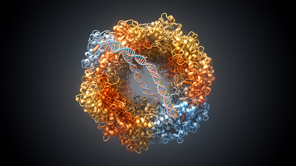
Deep inside every human cell, a molecular machine called the spliceosome performs one of the most intricate editing jobs in biology. It snips out non-coding stretches of RNA, known as introns, from freshly made messenger RNA precursors and stitches the remaining pieces together so that proteins can be built correctly. For decades, researchers have mapped how the spliceosome assembles and how it catalyses this cutting-and-pasting reaction in exquisite detail. Yet one crucial chapter of the story has remained stubbornly obscure: what happens after the reaction is finished, when the machine is left clutching the discarded intron and must somehow be taken apart so that its components can be recycled for the next round of splicing. A new study now provides the most complete structural picture yet of this final act, revealing a choreographed sequence of molecular events that researchers call spliceosome termination.
The spliceosome is a colossal ribonucleoprotein assembly, built from five small nuclear RNAs and well over a hundred proteins. It assembles anew on every single intron in every pre-messenger RNA molecule, and once it has excised the intron as a looped structure called a lariat, the ligated mRNA is released. But the excised intron-lariat stays wedged inside the RNA-based active site of the machine, forming a stable complex known as the intron-lariat spliceosome. This state is a dead end. To sustain the relentless pace of splicing in a growing cell, the intron-lariat spliceosome must be dismantled so that its proteins and small nuclear RNAs can be reused, and the trapped intron must be ejected, straightened out at its branch point, and ultimately degraded. These combined events constitute spliceosome termination, and until now the molecular details of how they unfold have been largely unknown.
A team led by researchers at the Research Institute of Molecular Pathology in Vienna, working with colleagues in the Netherlands and Spain, set out to catch the spliceosome in the act of terminating. Their strategy combined cryo-electron microscopy of spliceosomes purified directly from human cells with biochemistry, RNA sequencing and genetic experiments. By tagging the proteins DHX35 and YJU2B in human K562 cells and purifying the spliceosome complexes associated with them, the researchers obtained two distinct molecular snapshots. The first, resolved at a remarkable 2.4 angstrom resolution, captured a state they named the ILS-AQR complex. The second, at 3.2 angstroms, revealed an entirely new intermediate that they called the debranched intron spliceosome, or DIS. Together, these structures trace the sequential stages through which the spliceosome passes as it winds down its work.
The ILS-AQR structure revealed several surprises. The team identified a previously uncharacterized termination factor, YJU2B, bound to the spliceosome alongside the known termination factors TFIP11, C19L1, C19L2 and the RNA helicase DHX15. More strikingly, they found that the RNA helicase Aquarius, a protein already known for its role in spliceosome activation, was gripping the circular portion of the intron-lariat RNA in its active site. Aquarius belongs to a family of helicases that move along single-stranded RNA in a defined direction, and the structure showed it poised to pull on the intron-lariat branch point adenosine, the chemical knot that holds the lariat loop together. The researchers propose that Aquarius, guided by the RNA-binding protein PPIE, tugs the branch point out of the spliceosomal RNA network, while DHX15 simultaneously unwinds a different RNA element on the opposite side. Two RNAs pulled in opposite directions, the authors suggest, begin to unravel the intricate RNA architecture that holds the spliceosome together.
Extracting the branch point serves a second critical purpose: it exposes the knot so that it can be cut open. The enzyme responsible is DBR1, a debranching enzyme whose action is essential for intron turnover and for maintaining the balance of RNA in the cell nucleus. The new study provides the first molecular evidence that debranching happens directly on the spliceosome. Through a combination of proximity labelling, protein interaction predictions and biochemical validation, the researchers showed that DBR1 is tethered to the spliceosome through two conserved peptide segments in its tail. One of these peptides binds the termination factor C19L1, anchoring the catalytic domain of DBR1 within reach of the extracted branch point. Experiments in human cells confirmed the biological importance of this connection: DBR1 lacking the peptide could not rescue the growth defect caused by acute depletion of the endogenous enzyme, whereas the full-length protein could.
Once the branch point has been cut and the early termination factors have been displaced, the spliceosome enters the newly discovered DIS state. In this complex, the RNA network is almost completely dismantled. The U2 snRNP, the ESS2 protein and all six ILS-specific termination factors are gone, and the excised intron, now linearized, remains attached only through a short duplex between the U6 small nuclear RNA and the 5-prime splice site. RNA sequencing of the introns associated with each complex confirmed that the vast majority of DIS-bound introns had indeed been debranched, and that nearly all introns found in the ILS-AQR state were also detected in the DIS, indicating that passage through these two states is the predominant termination pathway for human introns.
The final blow is delivered by the RNA helicase DHX35, recruited to the DIS together with its cofactors GPATCH1 and WDR83. The structure shows DHX35 anchored on the spliceosome through extensive contacts with GPATCH1, which itself winds around the PRP8 protein at the heart of the machine. The U6 snRNA regions just downstream of the splice-site pairing are positioned to dock into the DHX35 active site, and the authors propose that DHX35 translocates along U6 snRNA, unwinding the remaining helical turn of the U6-intron duplex and ejecting the debranched intron for decay. In support of this model, purified DIS complexes immobilized on beads released their U5 snRNP and associated protein complexes upon addition of ATP, but only when DHX35 was catalytically competent. A mutant version of DHX35 unable to hydrolyse ATP bound the spliceosome normally yet failed to trigger disassembly, demonstrating that the helicase actively drives the final separation.
The study also illuminates how the cell deals with spliceosomes that go wrong. Splicing is not always successful, and defective complexes assembled on aberrant introns must be recognized and dismantled through a quality-control pathway. By revising an existing structure of a defective spliceosome from fission yeast, the researchers identified two additional factors, the YJU2B orthologue Saf4 and an uncharacterized protein called LENG1. LENG1, a paralogue of the essential branching factor CWC25, appears to partner with YJU2B specifically in defective complexes, where it helps guide the DHX35-GPATCH1-WDR83 machinery to the stalled spliceosome. The same helicases, DHX15 and DHX35, therefore serve both routine termination and quality control, but in regular termination they act in two sequential states, whereas in defective complexes they can be deployed simultaneously for a single-step disassembly.
The significance of this work extends beyond the mechanics of one molecular machine. Errors in splicing are implicated in a wide range of human diseases, including cancers driven by mutations in the splicing factor SF3B1, and the factors identified here, several of which rank among each other’s strongest genetic dependencies in human cell screens, may represent new points of vulnerability. By revealing how three RNA helicases, Aquarius, DHX15 and DHX35, orchestrate the gradual dismantling of the spliceosomal RNA network, coupling intron debranching and ejection to machine disassembly, the study closes a long-standing gap in our understanding of gene expression. Every protein-coding message in the human body depends on this final act of molecular housekeeping, and it has now, at last, been caught on camera.
Subject of Research: Structural mechanism of human spliceosome termination and intron-lariat debranching
Article Title: Mechanism of spliceosome termination
Article References: Boreikaite, V., Faraway, R., Vorländer, M. K., Phillips, A. W., Opitz, L., Wanke, M., Yakoub, G., Raffl, G., Fin, L., González-Prieto, R., Luijsterburg, M. S., Ameres, S. L., & Plaschka, C. (2026). Mechanism of spliceosome termination. Nature. https://doi.org/10.1038/s41586-026-11101-6
Image Credits: AI Generated
DOI: 10.1038/s41586-026-11101-6
Keywords: spliceosome, pre-mRNA splicing, cryo-electron microscopy, intron lariat, DHX35, DHX15, Aquarius, DBR1, RNA helicase, spliceosome recycling, RNA quality control, gene expression
News Source: Jason Bradley. (October 9, 2026). Scientists Capture the Spliceosome in the Act of Shutting Itself Down. Scienmag.