Lung adenocarcinoma is the most common form of non-small cell lung cancer, and while immune checkpoint blockade has transformed treatment for many patients, a substantial fraction of tumors either never respond or eventually stop responding to PD-1-targeting drugs. A study published in the Journal of Translational Medicine by Menglei Wang, Pengcheng Zhang, Xuefeng Li and colleagues now traces one reason why to an unexpected conversation between two stromal cell populations deep inside the tumor. The researchers report that a subset of macrophages marked by the protein SPP1 secretes the signaling molecule WNT5A, which switches on a population of myofibroblastic cancer-associated fibroblasts defined by the matrix metalloproteinase MMP11. These activated fibroblasts remodel the extracellular matrix, stiffen the tumor tissue, and physically and immunologically wall off the cancer from the CD8-positive T cells that immunotherapy relies upon.
The investigation began as a large-scale computational sweep. The team integrated bulk transcriptomic datasets, single-cell RNA sequencing and spatial transcriptomics from lung adenocarcinoma patients, cross-referencing these molecular maps with clinical cohorts that had received immune checkpoint blockade. Using weighted gene co-expression network analysis, survival meta-analysis and tissue enrichment scoring, they hunted for stromal cell subpopulations whose presence tracked with aggressive disease and poor immunotherapy outcomes. That search converged on a single culprit: a myofibroblastic cancer-associated fibroblast subset defined by high expression of MMP11, alongside the canonical contractile genes ACTA2 and TAGLN. These MMP11-positive myCAFs were enriched not only in primary lung tumors but also in metastatic lesions, and their abundance correlated with advanced tumor stage, metastatic spread, shorter overall survival and markedly reduced benefit from checkpoint inhibitors.
What makes this finding technically significant is the way the team moved beyond correlation. Spatial transcriptomic deconvolution, a computational method that infers which cell types occupy which physical niches within a tissue section, showed that MMP11-positive myCAFs consistently co-localized with SPP1-positive macrophages, a pro-tumor myeloid population that was itself enriched in malignant and metastatic lung adenocarcinoma tissue. The researchers then confirmed this spatial partnership in human clinical specimens using multiplex immunohistochemistry, a staining technique that visualizes several protein markers simultaneously on a single tissue slide. The two cell types were not merely neighbors in the same tumor; they occupied the same microanatomical neighborhoods, suggesting a functional relationship rather than coincidental distribution.
To identify the molecular language spoken between these cells, the team performed ligand-receptor interaction analysis on their single-cell and spatial data, complemented by virtual gene knockout experiments that computationally predicted the downstream consequences of removing specific signaling molecules. WNT5A emerged as the dominant macrophage-derived mediator linking SPP1-positive macrophages to myCAF activation. WNT5A is a member of the Wingless-related integration site family, a group of secreted glycoproteins that orchestrate embryonic development, tissue patterning and, in cancer, a bewildering array of pro-tumor behaviors. In this context, the analysis indicated that WNT5A released by macrophages acts on fibroblasts, driving them toward a matrix-remodeling, contractile state.
The mechanistic story has two halves. On the macrophage side, the researchers found that SPP1-positive macrophages exhibited activation of transcriptional programs governed by the non-canonical NF-kappaB factors NFKB2 and RELB. When they generated macrophages that overexpressed SPP1 in vitro, levels of Nfkb2, Relb and WNT5A protein all rose, placing SPP1 upstream of a signaling cascade that culminates in WNT5A production. This is a notable detail because it ties a well-known pro-tumor macrophage marker to a specific transcriptional pathway and a specific secreted output, giving the field a concrete molecular handle on a cell type that has often been described only by its marker genes.
On the fibroblast side, the team built a macrophage-CAF co-culture system in which SPP1-overexpressing macrophages and MMP11-overexpressing fibroblasts could interact directly. When fibroblasts were stimulated with WNT5A, they showed activation of YAP-associated signaling and increased expression of INHBA, the gene encoding the activin A subunit. YAP, the transcriptional co-activator downstream of the Hippo pathway, is a well-established sensor of mechanical forces and tissue stiffness, and its activation in fibroblasts is a hallmark of the myofibroblastic state. Functionally, WNT5A-stimulated fibroblasts migrated more readily, contracted more forcefully and deposited more extracellular matrix than their untreated counterparts. Each of these behaviors contributes to the desmoplastic reaction, the dense fibrous scar tissue that many solid tumors build around themselves.
The in vivo experiments brought the story into living systems. The researchers established murine tumor models and evaluated them with in vivo imaging, flow cytometry, atomic force microscopy, hematoxylin and eosin staining, Masson staining and multiplexed immunohistochemistry. Atomic force microscopy deserves particular mention: it measures tissue stiffness at the nanoscale by probing the physical resistance of the extracellular matrix, providing a direct readout of the mechanical remodeling that the fibroblasts were predicted to drive. The results were consistent with the in vitro findings. When WNT5A signaling was activated in the tumor microenvironment, collagen deposition increased, tissue stiffness rose, and MMP11-positive CAFs accumulated.
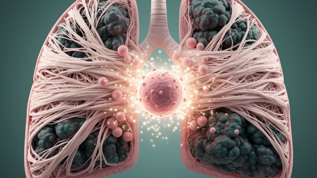
Most consequentially for patients, WNT5A activation impaired the therapeutic efficacy of PD-1 blockade in these animal models. Flow cytometry revealed reduced infiltration of CD8-positive T cells into the tumors, the very effector cells that anti-PD-1 antibodies are designed to unleash. The picture that emerges is a coherent immunosuppressive circuit: SPP1-positive macrophages, driven by NFKB2/RELB programs, secrete WNT5A; WNT5A activates YAP/INHBA signaling in fibroblasts; the fibroblasts remodel and stiffen the matrix; and the resulting dense, rigid stroma excludes cytotoxic T lymphocytes, leaving checkpoint inhibitors with little to work with even when the drug successfully engages its target on T cells.
The clinical implications are substantial. Immune checkpoint blockade has reshaped outcomes in lung adenocarcinoma, yet primary and acquired resistance remain stubborn problems, and stromal exclusion of T cells is one of the best-documented mechanisms of failure. By identifying SPP1-positive macrophages and MMP11-positive myCAFs as a spatially coupled, functionally linked pair, and by pinpointing WNT5A as the bridge between them, the study offers a set of potential biomarkers and drug targets. Patients whose tumors are rich in MMP11-positive myCAFs might be identified in advance as unlikely to benefit from PD-1 monotherapy and steered toward combination strategies. Meanwhile, the WNT5A-YAP axis suggests that disrupting macrophage-fibroblast communication could soften the tumor matrix, restore T-cell infiltration and resensitize tumors to immunotherapy. The authors note that their findings highlight a potential therapeutic strategy to overcome immune checkpoint blockade resistance, though translating that insight into clinics will require inhibitors capable of safely modulating WNT signaling in humans.
Methodologically, the study is also a demonstration of how modern cancer biology is increasingly conducted. No single technique carried the argument; instead, bulk transcriptomics established population-level associations, single-cell RNA sequencing resolved the relevant cell states, spatial transcriptomics and multiplex immunohistochemistry anchored those states in physical tissue architecture, virtual knockout and co-culture experiments tested causal mechanisms in vitro, and mouse models with mechanical measurements confirmed the circuit in vivo. The human specimens were obtained under ethics approval from Zhejiang Cancer Hospital, and the work was supported by the National Natural Science Foundation of China and the Traditional Chinese Medicine Scientific Research Foundation of Zhejiang Province. As with any study that combines computational inference with experimental validation, the next step will be independent confirmation in larger immunotherapy cohorts and the development of clinically tractable ways to interrupt the WNT5A signal. If those hurdles can be cleared, the stromal conversation between macrophages and fibroblasts may become one more vulnerable link in the armor that lung adenocarcinomas use to shield themselves from the immune system.
Subject of Research: The role of SPP1-positive macrophage-derived WNT5A in MMP11-positive myofibroblastic cancer-associated fibroblast activation, matrix remodeling, and immunotherapy resistance in lung adenocarcinoma.
Article Title: SPP1⁺ macrophage-derived WNT5A contributes to MMP11⁺ myCAF-mediated matrix remodeling and immunotherapy resistance in lung adenocarcinoma
Article References: Wang, M., Zhang, P., Li, X., Shao, L., Dai, S., & Chen, Z. (2026). SPP1⁺ macrophage-derived WNT5A contributes to MMP11⁺ myCAF-mediated matrix remodeling and immunotherapy resistance in lung adenocarcinoma. Journal of Translational Medicine. https://doi.org/10.1186/s12967-026-09069-y
Image Credits: AI Generated
DOI: 10.1186/s12967-026-09069-y
Keywords: lung adenocarcinoma, WNT5A, SPP1 macrophages, cancer-associated fibroblasts, MMP11, matrix remodeling, immunotherapy resistance, PD-1 blockade, tumor microenvironment, YAP signaling, single-cell RNA sequencing, spatial transcriptomics
News Source: Nathaniel Bowman. (October 9, 2026). Macrophage Signal WNT5A Drives Lung Cancer Scarring That Blocks Immunotherapy. Scienmag.