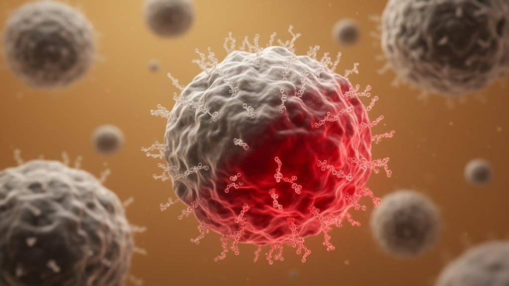
Macrophages are among the most versatile sentinels of the human immune system. In their normal state, these white blood cells engulf bacteria, clear cellular debris, and mount aggressive attacks against emerging cancer cells. But when macrophages infiltrate a growing tumor, something changes. They become tumor-associated macrophages, or TAMs, and many of them gradually abandon their inflammatory, tumor-fighting behavior in favor of an anti-inflammatory state that dampens the immune response. In the context of cancer, that shift is dangerous: a suppressed immune system gives tumors room to grow unchecked, and colorectal cancer patients whose tumors harbor high levels of these anti-inflammatory TAMs tend to fare worse. Now, an international research team has identified a molecular mechanism that appears to drive this transformation, and, more strikingly, has shown in laboratory experiments that blocking a single enzyme can push the cells back toward their cancer-fighting identity.
The study, led by Assistant Professor Priya Dipta of Nagoya University’s Institute for Glyco-Core Research in Japan and Professor Morten Thaysen-Andersen of Macquarie University in Australia, with collaborators including Daisuke Kasugai, Hironoshin Onizuka and colleagues at Nagoya University Graduate School of Medicine, was published in the Proceedings of the National Academy of Sciences of the United States of America. The team’s findings center on a layer of the cell surface that has long been overlooked in cancer immunology: the glycocalyx, a dense coating of proteins and lipids studded with sugar chains that mediates how cells communicate with one another and with their surroundings.
The sugar chains of the glycocalyx are frequently capped with sialic acid, a negatively charged sugar that influences everything from cell adhesion to immune recognition. Sialic acids attach to underlying glycans through two main linkage types, designated α2,3 and α2,6. Altered sialylation is already known to help cancer cells evade immune surveillance, but how these linkages change as macrophages shift between pro-inflammatory and anti-inflammatory states had remained unclear. To answer that question, the researchers isolated monocytes from healthy donors and differentiated them in the laboratory into tumor-fighting, pro-inflammatory macrophages and tumor-promoting, anti-inflammatory macrophages, then compared the sialic acid linkages displayed on their surfaces.
The contrast was dramatic. Using mass spectrometry and glycan-specific protein staining, the team found that tumor-fighting macrophages carried predominantly α2,3-linked sialic acids on their surface N-glycans. In tumor-promoting macrophages, the situation was nearly reversed: roughly ninety percent of their sialic acids were attached through the α2,6 linkage. This linkage switch, the researchers concluded, is a hallmark of the macrophage transition from a state that attacks tumors to one that tolerates and even supports them.
Perhaps the most visually striking discovery came from fluorescence microscopy. The tumor-promoting macrophages extended long, branching protrusions coated with α2,6-linked sialic acid, which the researchers named sialo-protrusions. These structures reached up to 800 micrometers from the cell body, an extraordinary distance for cellular extensions, and formed mesh-like networks by connecting neighboring cells. Tumor-fighting macrophages displayed far fewer of these structures. Intriguingly, the sialo-protrusions showed no evidence of actin, the protein that typically forms cellular extensions such as filopodia and lamellipodia. Instead, they carried α2,6-sialylated glycoproteins on their surface, suggesting they represent a previously unrecognized class of cellular projections distinct from known actin-based structures.
The search for the molecular switch behind this remodeling led the team to an enzyme called ST6GAL1, which catalyzes the addition of sialic acid in the α2,6 configuration. Only the tumor-promoting macrophages expressed high levels of ST6GAL1, making it a prime suspect in the linkage switch. When the researchers silenced the enzyme using siRNA, the effects were immediate and widespread. The sialo-protrusions broke apart and shortened, the macrophages moved less, and they interacted less with one another, indicating that these sugar-coated extensions are functionally important for macrophage motility and communication rather than mere decoration.
The consequences extended to the cancer cells themselves. With ST6GAL1 suppressed, the tumor-promoting macrophages made less contact with colorectal cancer cells in laboratory co-cultures, and the cancer cells grew much more slowly. This suggests that sialo-protrusions help macrophages migrate, communicate with cancer cells, and support tumor growth through those physical contacts. Removing the enzyme that builds the α2,6 linkages appears to sever a critical line of communication between immunosuppressive macrophages and the tumor.
Even more remarkably, suppressing ST6GAL1 did more than disable the protrusions. The treated tumor-promoting macrophages began to resemble their tumor-fighting counterparts in shape, in the cytokines they expressed, and in their phagocytic ability, the capacity to engulf targets. In other words, targeting a single enzyme appeared to shift the cells’ overall state back toward the pro-inflammatory, tumor-attacking program. The finding raises the possibility that glycocalyx remodeling could become a therapeutic lever for reprogramming the immune microenvironment of tumors rather than simply blocking one interaction at a time.
To confirm that the laboratory observations reflected biology in real patients, the team examined tumor tissue from patients with advanced colorectal cancer. The same pattern held: tumor-promoting TAMs carried more α2,6-linked sialic acid, while tumor-fighting TAMs displayed more α2,3 linkages, mirroring the results from the donor-derived cells. This consistency between controlled experiments and patient samples strengthens the case that the sialyl linkage switch is a genuine feature of the tumor immune landscape rather than an artifact of cell culture. Dipta noted that the findings provide a new perspective on how remodeling of the immune cell glycocalyx may influence the tumor microenvironment.
Important questions remain before the discovery can be translated into treatment. The researchers emphasize that future studies must test ST6GAL1 inhibition in living organisms to evaluate whether it produces meaningful antitumor effects in the complex setting of a real tumor, where macrophages interact with many other immune and stromal cell types. The team also aims to identify the proteins that form the sialo-protrusions and to clarify exactly how these unusual structures mediate communication between macrophages and cancer cells. If those efforts succeed, they could open a new strategy for cancer immunotherapy: not boosting immune cells with checkpoint blockers alone, but chemically re-sculpting the sugar coat that tells macrophages which side to fight for. For now, the study offers a compelling proof of concept that the fate of tumor-associated macrophages may hinge, quite literally, on the fine chemical details of the sugars they display.
Subject of Research: Glycocalyx sialylation and macrophage polarization in the colorectal tumor microenvironment
Article Title: Shifting immune cells back toward cancer-fighting mode
Article References: Shifting immune cells back toward cancer-fighting mode. (n.d.). Original publication
Image Credits: AI Generated
DOI: Not provided
Keywords: macrophages, tumor-associated macrophages, glycocalyx, sialic acid, ST6GAL1, colorectal cancer, tumor microenvironment, sialo-protrusions, glycosylation, cancer immunotherapy, Nagoya University, PNAS
News Source: Nathaniel Bowman. (October 8, 2026). Sugar Switch on Immune Cells Could Push Macrophages Back Into Cancer-Fighting Mode. Scienmag.