Scientists at the University of Oklahoma have identified three small subclusters of cells that work together to drive cachexia, the devastating muscle-wasting and fat-loss syndrome that afflicts the majority of patients with pancreatic cancer. The research, published in the journal Cell, reveals that these subclusters—SEMA4A-positive tumor cells, AQP9-positive macrophages and LOXL2-positive cancer-associated fibroblasts—form a physically adjacent microenvironment that behaves like a self-reinforcing engine of tissue wasting. The discovery, led by senior author Min Li, Ph.D., professor of medicine in the OU College of Medicine and associate director for global oncology at OU Health Stephenson Cancer Center, offers the most detailed spatial map to date of how a tumor creates the conditions for its host to deteriorate, and it points directly toward new therapeutic strategies for a condition that currently has no effective treatment.
Cachexia is far more than simple weight loss. It is a systemic syndrome characterized by the progressive loss of skeletal muscle and adipose tissue that cannot be reversed by nutritional support alone. Patients become weak, lose appetite and strength, and—critically—become less able to tolerate chemotherapy, targeted therapy or surgery. In pancreatic cancer, the problem is especially severe: more than 80 percent of patients develop cachexia, and the disease carries a five-year survival rate below 13 percent across all stages. Because treatment tolerance is so tightly linked to outcomes, a patient who becomes cachexic early often cannot receive full therapeutic doses, creating a vicious cycle in which the tumor advances while the body’s defenses collapse. Understanding the biological triggers of this cycle has therefore been one of the most urgent unmet needs in oncology.
To find those triggers, Li’s team deployed cutting-edge technologies capable of resolving tumors at extraordinary resolution. Single-cell sequencing allowed the researchers to separate the tumor microenvironment into its individual cellular components, while spatial transcriptomics revealed where each of those components sat in relation to its neighbors. This spatial dimension proved decisive. The three subclusters were not scattered randomly through the tumor; they were physically adjacent to one another, forming what the researchers call a molecular niche—a localized microenvironment whose architecture is conducive to the initiation and progression of cachexia. The finding suggests that cachexia is not simply the product of circulating factors released by tumors into the bloodstream, but of a specific, spatially organized cellular consortium operating within the tumor itself.
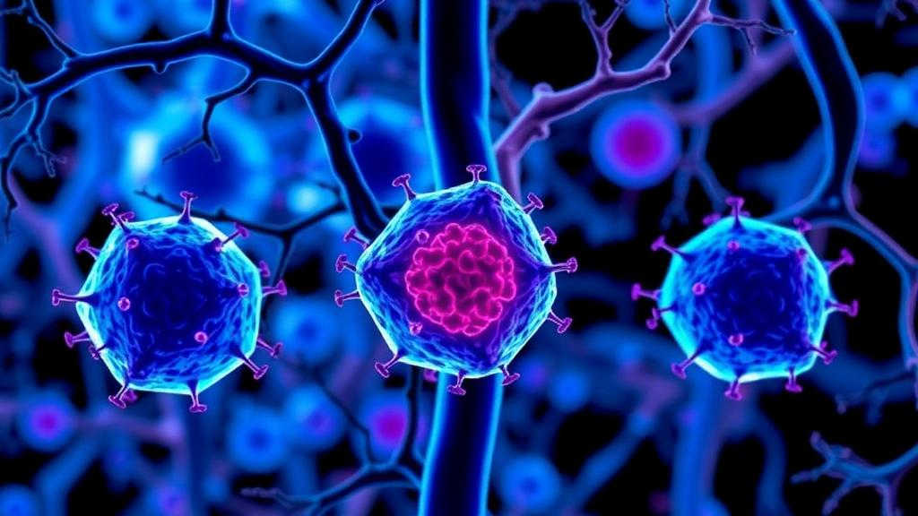
Equally striking is the way the three subclusters interact. Together they form a triangular regulatory network operating in a feed-forward loop, meaning each component amplifies the activity of the others, accelerating the onset and progression of wasting over time. This architecture helps explain why cachexia, once established, has proven so resistant to intervention: targeting any single player may leave the other two arms of the triangle intact. The identification of all three participants simultaneously—the SEMA4A-expressing tumor cells, the AQP9-expressing macrophages and the LOXL2-expressing fibroblasts—provides, for the first time, a complete set of targets whose combined disruption could theoretically break the loop.
These are the players that are driving cachexia in pancreatic cancer patients, Li explained. His team successfully identified and isolated the small cell clusters from each cell type in the tumor microenvironment, and the next step, he said, is to develop specific strategies to target the three molecules that define them. Such strategies could take the form of inhibitors, antibodies or other agents designed to neutralize the signaling that flows among the subclusters. Because the subclusters are defined by specific molecular markers, they also serve as potential biomarkers: their presence in a tumor may help clinicians identify which patients are likely to progress to pre-cachexia and full cachexia, enabling intervention before irreversible tissue loss begins.
That early-detection angle may prove as important as the therapeutic one. Li emphasized that the subclusters form before muscle and fat loss actually occur, meaning the window for preventive treatment exists if physicians know what to look for. Addressing cachexia early could ultimately help patients remain strong enough to tolerate the full course of their cancer treatment. Any eventual anti-cachexia therapy, however, would need to be delivered in combination with the cancer treatment itself, whether chemotherapy or a targeted agent. We need to slow down tumor growth at the same time we’re slowing the progression of cachexia, Li said. Otherwise, if clinicians only lower the tumor burden, patients quickly become cachexic, lose muscle strength and appetite, and become less able to withstand treatment.
The publication is the culmination of a multi-year research arc in which Li’s laboratory has progressively refined the field’s understanding of how wasting begins. In a 2024 paper in Cancer Cell, his team discovered that crosstalk between pancreatic cancer cells and macrophages—the immune cells that populate tumors—is the first step toward the onset of cachexia. Earlier this year, in a second Cancer Cell paper, he articulated a triangle regulation theory in which cancer cells recruit and activate macrophages, which in turn enlist the involvement of the central nervous system. The new Cell study extends that framework from a two-way conversation into a fully mapped three-party niche, adding cancer-associated fibroblasts as a third partner and pinpointing the specific molecular subtypes of each cell that matter.
Leadership at the University of Oklahoma framed the work as a milestone for both science and patient care. Robert S. Mannel, M.D., director of the OU Health Stephenson Cancer Center, said cachexia has a profound impact on patients’ quality of life and their ability to tolerate cancer treatment, and that the discovery gives researchers new insight into what drives the syndrome while opening the door to detecting and treating it earlier. Ian F. Dunn, M.D., executive dean of the OU College of Medicine and chief physician executive for OU Health, said the study demonstrates how fundamental scientific discovery can lay the groundwork for advances in patient care, describing the findings as a foundation for new therapeutic strategies against a debilitating condition and noting that publication in Cell reflects the significance of the work and the caliber of discovery taking place at the OU College of Medicine on the Harold Hamm Health Campus.
The study, titled Spatial Evolution of a Cachexia-Promoting Microenvironment in Pancreatic Cancer, was supported by the OU College of Medicine, the college’s Department of Medicine and the OU Health Stephenson Cancer Center, which is supported in part by Oklahoma’s Tobacco Settlement Endowment Trust. For patients and clinicians, the significance is hard to overstate. There are no good treatments for cachexia today, Li noted, which is precisely why the identification of a defined, targetable cellular network is so exciting. If therapies can be developed against the SEMA4A, AQP9 and LOXL2 subclusters—and if those subclusters can serve as early warning markers in diagnostic testing—the field may finally gain the tools to intervene before wasting begins rather than after it has taken hold. For a disease in which most patients develop cachexia and nutrition alone cannot reverse it, converting a fatal complication into a manageable one would represent a genuine turning point in pancreatic cancer care.
Subject of Research: Cellular mechanisms of pancreatic cancer-induced cachexia
Article Title: Cell paper identifies subclusters driving cancer-induced cachexia
Article References: Cell paper identifies subclusters driving cancer-induced cachexia. (n.d.). Original publication
Image Credits: AI Generated
DOI: Not provided
Keywords: pancreatic cancer, cachexia, tumor microenvironment, single-cell sequencing, spatial transcriptomics, macrophages, cancer-associated fibroblasts, molecular niche, SEMA4A, AQP9, LOXL2, Cell journal
News Source: Nathaniel Bowman. (October 6, 2026). Three Cell Subclusters Uncovered as Drivers of Pancreatic Cancer Cachexia. Scienmag.