Credit: Phil Mcleod/Michigan Tech
Get your popcorn. Engineers and virologists have a new way to watch viral infection go down.
The technique uses microfluidics — the submillimeter control of fluids within a precise, geometric structure. On what is basically a tricked-out microscope slide, chemical engineers from Michigan Technological University have been able to manipulate viruses in a microfluidic device using electric fields. The study, published this summer in Langmuir, looks at changes in the cell membrane and gives researchers a clearer idea of how antivirals work in a cell to stop the spread of infection.
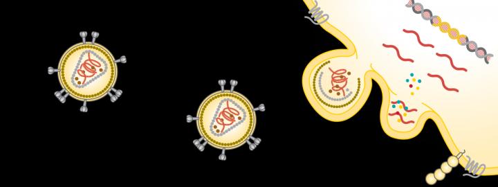
Viruses carry around an outer shell of proteins called a capsid. The proteins act like a lockpick, attaching to and prying open a cell’s membrane. The virus then hijacks the cell’s inner workings, forcing it to mass produce the virus’s genetic material and construct many, many viral replicas. Much like popcorn kernels pushing away the lid of an overfilled pot, the new viruses explode through the cell wall. And the cycle continues with more virus lockpicks on the loose.
“When you look at traditional techniques — fluorescent labeling for different stages, imaging, checking viability — the point is to know when the membrane is compromised,” said Adrienne Minerick, study co-author, dean of the College of Computing and a professor of chemical engineering. “The problem is that these techniques are an indirect measure. Our tools look at charge distribution, so it’s heavily focused on what’s happening between the cell membrane and virus surface. We discovered with greater resolution when the virus actually goes into the cell.”
Watching the viral infection cycle and monitoring its stages is crucial for developing new antiviral drugs and gaining better understanding of how a virus spreads. Dielectrophoresis happens when polarizable cells get pushed around in a nonuniform electric field. The movement of these cells is handy for diagnosing diseases, blood typing, studying cancer and many other biomedical applications. When applied to studying viral infection, it’s important to note that viruses have a surface charge, so within the confined space in a microfluidic device, dielectrophoresis reveals the electric conversation between the virus capsid and the proteins of a cell membrane.
“We studied the interaction between the virus and cell in relation to time using microfluidic devices,” said Sanaz Habibi, who led the study as a doctoral student in chemical engineering at Michigan Tech. “We showed we could see time-dependent virus-cell interactions in the electric field.”
Watching a viral infection happen in real time is like a cross between a zombie horror film, paint drying and a Bollywood epic on repeat. The cells in the microfluidic device dance around, shifting into distinct patterns with a dielectric music cue. There needs to be the right ratio of virus to cells to watch infection happen — and it doesn’t happen quickly. Habibi’s experiment runs in 10-hour shifts, following the opening scenes of viral attachment, a long interlude of intrusion, and eventually the tragic finale when the new viruses burst out, destroying the cell in the process.
Before they burst, cell membranes form structures called blebs, which change the electric signal measured in the microfluidic device. That means the dielectrophoresis measurements grant high-resolution understanding of the electric shifts happening at the surface of the cell through the whole cycle.
Viral infections are top of mind right now, but not all viruses are the same. While microfluidic devices that use dielectrophoresis could one day be used for on-site, quick testing for viral diseases like COVID-19, the Michigan Tech team focused on a well-known and closely studied virus, the porcine parvovirus (PPV), which infects kidney cells in pigs.
But then the team wanted to push the envelope: They added the osmolyte glycine, an important intervention their collaborators study in viral surface chemistry and vaccine development.
“Using our system, we could show time-dependent behavior of the virus and cell membrane. Then we added the osmolyte, which can act as an antiviral compound,” Habibi explained. “We thought it would stop the interaction. Instead, it looked like the interaction continued to happen at first, but then the new viruses couldn’t get out of the cell.”
That’s because glycine likely interrupts the new capsid formation for the replicated viruses within the cell itself. While that specific portion of the viral dance happens behind the curtain of the cell wall, the dielectric measurements show a shift between an infected cycle where capsid formation happens and an infected cell where capsid formation is interrupted by glycine. This difference in electrical charge indicates that glycine prevents the new viruses from forming capsids and stops the would-be viral lockpickers from hitting their targets.
“When you are working with such small particles and organisms, when you’re able to see this process happening in real time, it’s rewarding to track those changes,” Habibi said.
This new view of the interactions between virus capsids and cell membranes could speed up testing and characterizing viruses, cutting out expensive and time-consuming imaging technology. Perhaps in a future pandemic, there will be point-of-care, handheld devices to diagnose viral infections and we can hope medical labs will be outfitted with other microfluidic devices that can quickly screen and reveal the most effective antiviral medications.
###
Media Contact
Allison Mills
[email protected]
Original Source
https:/
Related Journal Article
http://dx.


