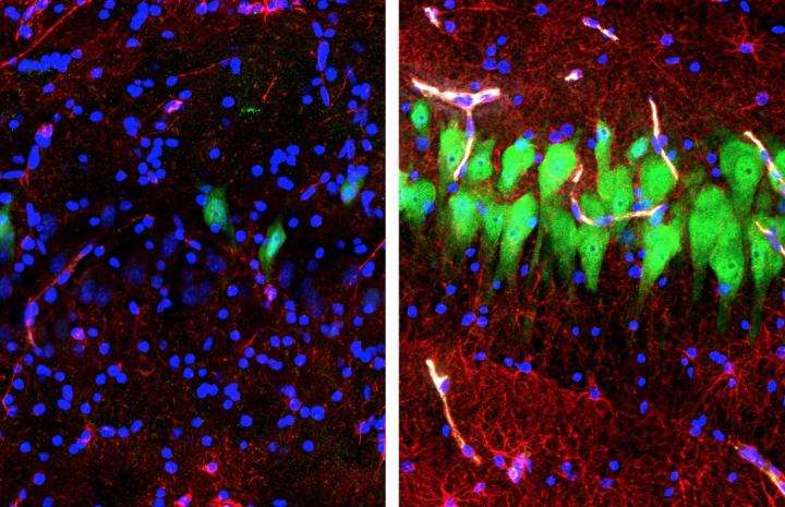
Tissue support system preserves limited function in an isolated postmortem animal brain

Credit: Stefano G. Daniele & Zvonimir Vrselja; Sestan Laboratory; Yale School of Medicine
Researchers have developed a high-tech support system that can keep a large mammalian brain from rapidly decomposing in the hours after death, enabling study of certain molecular and cellular functions. With funding through the National Institutes of Health BRAIN Initiative, researchers developed a way to deliver an artificial blood to the isolated postmortem brain of a pig, preventing the degradation that would otherwise destroy many cellular and molecular functions and render it unsuitable for study. Importantly, although the researchers saw some preservation of flow through blood vessels and energy use, there was no functional activity in the brain circuits. The scientific team, led by Nenad Sestan, M.D., Ph.D., of Yale University, New Haven, Connecticut, reports on their findings in the journal Nature.
“This line of research could lead to a whole new way of studying the postmortem brain,” explained Andrea Beckel-Mitchener, Ph.D., BRAIN Initiative Team Lead at the NIH’s National Institute of Mental Health, which co-funded the research. “The new technology opens up opportunities to examine complex cell and circuit connections and functions that are lost when specimens are preserved in other ways. It also could stimulate research to develop interventions that promote brain recovery after loss of brain blood flow, such as during a heart attack.”
Researchers’ ability to study the functional dynamics of an intact, isolated large postmortem brain has been hampered by cell death, blockage of small blood vessels and other toxic processes that degrade the tissue following loss of blood flow and oxygen. Freezing and other preservation methods allow for only static microscopic, biochemical or structural analyses.
To overcome these limitations, Sestan and colleagues created a system called BrainEx (after “ex vivo”), specially designed to attenuate some of the processes responsible for degradation of tissue in postmortem brains. The researchers used brains from a pork processing plant that would have otherwise been discarded. The system involves pumping a solution called BEx perfusate – a proprietary mixture of protective, stabilizing and contrast agents, that act as substitutes for blood – into the isolated brain’s main arteries at normal body temperature.
Brains processed with BEx showed reduced cell death, preserved anatomical and cell architecture, restored blood vessel structure and circulatory function, restored glial inflammatory responses, spontaneous neural activity at synapses and active cerebral metabolism, compared to brains perfused with a control solution, which rapidly decomposed. There was no global electrical activity that would indicate higher-order functions, such as awareness or perception.
The results suggest that delivering protective agents to the brain through its dense network of blood vessels may hold potential for improving survival and reducing neurological deficits after trauma.
Among potential future applications, the researchers suggest BrainEx might be used to test how an experimental drug affects the intricate 3-D wiring of a large brain. The BEx system may also help researchers use postmortem brain specimens to study the effects of brain injury on cells and neural connections.
“BrainEx’s cell-protective formulations may someday find application in therapies for disorders such as stroke,” said Sestan. “The isolated large mammalian brain’s capacity for restoration of microcirculatory, molecular and cellular activity has been underappreciated.”
The BRAIN Initiative was founded on the principle that new tools and advanced technologies are needed to accelerate scientific discoveries that can lead to a better understanding of brain function and human diseases. While the researchers stress that BrainEx offers ample opportunities for scientific exploration in animal postmortem brains without restoration of brain circuit activity, they specify contingencies that would guard against crossing of ethical lines should any signs of such functions appear as the technology develops.
“As medical researchers, we have an ethical imperative to use the powerful tools developed by BRAIN Initiative researchers to help unravel the mysteries of brain injuries and diseases,” said Christine Grady, R.N., Ph.D., chief of the Department of Bioethics at the NIH Clinical Center. “It’s also our duty to work with researchers to thoughtfully and proactively navigate any potential ethical issues they may encounter as they open new frontiers in brain science. Since the launch of the BRAIN Initiative in 2013, NIH has been working with experts to address these questions.”
The BRAIN Initiative is funding a growing network of neuroethics researchers. NIH has also been working with experts, including members of the NIH BRAIN Initiative Neuroethics Working Groupreports toreof, to help guide the integration of neuroethics across projects as our understanding and capabilities expand. NIH recently published an overview of its multi-faceted and proactive approach to navigate potential ethical implications of new technologies funded by the BRAIN Initiative. A keystone of the NIH BRAIN Initiative’s neuroethics strategy is a set of Neuroethics Guiding Principles. Further, the NIH, in conjunction with the Neuroethics Working Group, organizes workshops to explore emerging areas of science that raise ethical questions, including one last year that focused on research with human neural tissue
###
Reference:
Restoration of brain circulation and cellular functions hours postmortem. Vrselja Z, Daniele SG, Silbereis J, Talpo F, Morozov YM, Sousa AMM, Tanaka BS, Skarica M, Pletikos M, Kaur N, Zhuang ZW, Liu Z, Alkawadri R, Sinusas AJ, Latham SR, Waxman SG, Sestan N. Nature, April 18, 2019.
About the National Institute of Mental Health (NIMH): The mission of the NIMH is to transform the understanding and treatment of mental illnesses through basic and clinical research, paving the way for prevention, recovery and cure. For more information, visit the NIMH website.
The National Institute of Neurological Disorders and Stroke (NINDS) is the nation’s leading funder of research on the brain and nervous system. The mission of NINDS is to seek fundamental knowledge about the brain and nervous system and to use that knowledge to reduce the burden of neurological disease.
The National Institute of General Medical Sciences (NIGMS) is a part of the National Institutes of Health that supports basic research to increase our understanding of biological processes and lay the foundation for advances in disease diagnosis, treatment, and prevention. For more information on the Institute’s research and training programs, visit http://www.
About the NIH Clinical Center: The NIH Clinical Center is the clinical research hospital for the National Institutes of Health. Through clinical research, clinician-investigators translate laboratory discoveries into better treatments, therapies and interventions to improve the nation’s health. More information: https:/
About the National Institutes of Health (NIH): NIH, the nation’s medical research agency, includes 27 Institutes and Centers and is a component of the U.S. Department of Health and Human Services. NIH is the primary federal agency conducting and supporting basic, clinical, and translational medical research, and is investigating the causes, treatments, and cures for both common and rare diseases. For more information about NIH and its programs, visit the NIH website.
BrainEx Fact Sheet
Overview:
- With funding through the National Institutes of Health (NIH) BRAIN Initiative, researchers at Yale University developed a high-tech support system that can preserve and protect an isolated mammalian brain from breakdown in the hours after death, which may transform how we study brain structure and function.
- The system, called BrainEx, artificially pulsed a cell-protective solution through the blood vessels of an isolated postmortem pig brain. The technique prevented degradation that would otherwise destroy many cellular and molecular functions and thus preserved several aspects of brain structure and chemistry to facilitate the scientific study of its properties.
Background:
- Mammalian brains have high energy demands, making them susceptible to damage after a stoppage of blood flow.
- Unless blood flow is quickly restored to the brain, a cascade of cell damage and death –
as well as tissue destruction – occurs, known as brain death.In addition to cellular death, multiple mechanisms lead to widespread obstruction of flow through the small vessels so that brain blood flow can no longer be restored. - Prior studies of mammalian brain and brain tissue called into question just how fast this rapid brain degradation follows death, hinting that certain aspects of brain structure and cellular activity could be maintained for a small window of time during the postmortem period, or after prolonged ischemic events.
- This led the researchers to examine whether, under appropriate conditions, certain brain cells could be protected from degrading so quickly after blood flow ceases.
About the Study:
Methods:
- The researchers developed a pulsatile perfusion system, BrainEx (BEx) which uses a combination of pumps to deliver a proprietary cell-protective solution to a large mammalian postmortem brain through its vascular system.
- The postmortem pig brains used in the study were sourced from a pork processing plant and would have been otherwise discarded.
- Four hours after death the brain was connected to the BrainEx system for 6 hours and a hemoglobin-based, acellular, echogenic and non-coagulative solution called BEx perfusate – comprising various cell-protective compounds, including blockers of neuronal activity – was pumped through the isolated brain’s blood vessels at normal body temperature.
- The researchers employed various independent imaging methods as well as careful studies at the tissue and ultrastructural level across multiple brain regions to analyze the extent to which anatomical and cellular integrity was maintained under BEx conditions.
Results:
- Compared to non-perfused or brains perfused with the control solution, BrainEx-treated brains showed:
- o reduced cell death;
o preserved anatomical and cell architecture;
o restored blood vessel structure and filling of large and small blood vessels.
o restored glial inflammatory responses;
o active cerebral metabolism of glucose and oxygen;
o in vitro spontaneous neural activity at synapses in cells that were removed from the BrainEx-treated brains and studied under the microscope.
- Brains connected to the BrainEx system did not show any global electrical activity consistent with normal brain function.
- Brains that were not perfused with the BrainEx solution rapidly decomposed in all parameters measured.
Implications:
- The rapid cell damage that occurs after brain death has led researchers to preserve brains through methods such as quick freezing them or “fixing” them with formaldehyde — however, these processes make the brains unusable for many types of scientific research, such as assessing cellular functions and connections between nerve cells.
- These findings provide a new avenue for research that is currently impossible to accomplish, due to both drawbacks of current brain-preservation techniques and experimental and ethical limitations in applying classical molecular techniques to the in vivo large mammalian brain.
- The authors note that the observed restoration of limited molecular and cellular processes should not be interpreted to indicate resurgence of normal brain function.
- Brain death is defined as the irreversible loss of all functions of the brain, including the brainstem, but does not imply all brain cells are dead at the time the diagnosis is made.
- The study could spur research to develop more effective means of protecting the brain after catastrophic injury due to conditions such as cardiac arrest.
- Although the researchers continuously monitored the brains for signs of functional activity, at no point did they observe the kind of organized global electrical activity associated with awareness, perception or any other higher-order brain functions.
The NIH BRAIN Initiative
- Launched in 2013, the Brain Research through Advancing Innovative Neurotechnologies® (BRAIN) Initiative aims to accelerate neuroscience research by equipping scientists with new tools to improve our understanding of the brain and its disorders, including Alzheimer’s disease, schizophrenia, autism, epilepsy and traumatic brain injury.
- With support from Congress through both the regular appropriations process and the 21st Century Cures Act, the NIH’s BRAIN Initiative has funded over 550 awards, totaling more than $900 million, through fiscal year 2018.
- A goal of the NIH’s BRAIN Initiative is to develop and apply innovative neurotechnologies to understand how brain circuits work to enable cognition, emotion, perception, action and what goes wrong in disease.
NIH BRAIN Initiative Ethical Issues and Solutions
- Finding new and innovative ways for scientists to study the human brain is essential for developing better treatments for the brain diseases and disorders that afflict millions in the United States and worldwide, including depression, chronic pain, Alzheimer’s disease and substance use disorders.
- The emergence of new technologies may require an examination of ethical standards to guide how those technologies should be introduced into scientific and clinical practice.
- NIH emphasizes integrating neuroethics throughout the BRAIN Initiative, via proactive, ongoing assessment of the neuroethical implications of the development and application of BRAIN-funded tools and neurotechnologies. The NIH BRAIN Initiative also supports a portfolio of neuroethics research, examining the ethical implications of advances in neurotechnology and brain science.
- In 2015, the NIH BRAIN Initiative established its Neuroethics Working Group, composed of 10 experts in neuroethics and neuroscience.
- The group serves to help ensure neuroethics is integrated throughout the NIH BRAIN Initiative by:
- o reviewing the portfolio of BRAIN grants and identifying areas of research that raise ethical questions;
o discussing those ethical questions, in conjunction with the relevant BRAIN Initiative investigators and invited ethics experts;
o providing consultations on specific projects; and
o recommending neuroethics questions that are amenable to research.
- The BRAIN Initiative’s Neuroethics Working Group developed a set of Neuroethics Guiding Principles for the NIH BRAIN Initiative that were, alongside a commentary from the NIH BRAIN Initiative Institute and Center Directors, published in the Journal of Neuroscience in December 2018.
- The Neuroethics Working Group provides help to organize topical workshops that explore neuroethical implications of BRAIN Initiative research; topics to date have included human neuroscience research utilizing invasive and noninvasive neural devices, and research with human neural tissue.
- The Neuroethics Working Group also is available to provide consultations for individual BRAIN investigators. On the recommendation of his NIH program officer, in 2016 Dr. Sestan engaged in a consultation with the Neuroethics Working Group on BrainEx.
- o This subsequently led to two topical workshops on research with human neural tissue, supported by the NIH BRAIN Initiative.
o At the most recent of these workshops in March of 2018, a key part of the conversation focused on continuing to ensure that research working with invaluable ex vivo human brain tissue is done in a responsible manner and adheres to the highest ethical standards.
Media Contact
Jules Asher
[email protected]


