Credit: The University of Edinburgh
Scientists have developed a new imaging technology to visualise what cells eat, which could aid the diagnosis and treatment of diseases such as cancer.
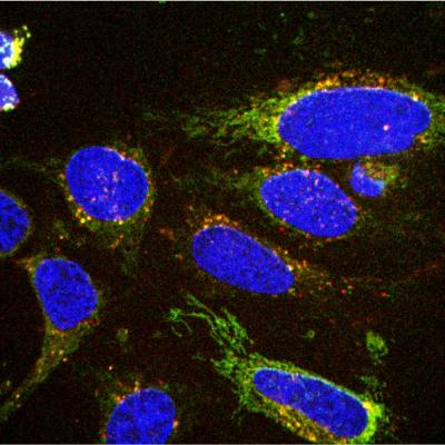
The team has designed chemical probes that light up when they attach to specific molecules that cells eat, such as glucose.
Researchers used microscopes to watch cells eating glucose inside live zebrafish embryos, which are transparent and easy to observe. They found the technique also worked with human cells growing in the lab.
The team says their approach could easily be adapted to look at other molecules that are important for health and disease.
All cells rely on glucose and other molecules for their survival. If a cell’s eating habits change, it can be a warning sign of disease.
Researchers say the new technology could help detect tiny changes in cells’ eating habits inside the body’s tissues, making it easier to spot diseases sooner.
Doctors could also use the technology to monitor how patients are responding to treatment, by tracking the molecules that are eaten by healthy and diseased cells.
The study, published in the journal Angewandte Chemie, was funded by Medical Research Scotland, the Biotechnology and Biological Sciences Research Council and the European Research Council. The Royal Society and the Wellcome Trust also provided funding.
Dr Marc Vendrell, Senior Lecturer in Biomedical Imaging at the University of Edinburgh, said: “We have very few methods to measure what cells eat to produce energy, which is what we know as cell metabolism. Our technology allows us to detect multiple metabolites simultaneously and in live cells, by simply using microscopes.
“This is a very important advance to understand the metabolism of diseased cells and we hope it will help develop better therapies.”
###
Media Contact
Jen Middleton
[email protected]
Related Journal Article
http://dx.


