A critical narrative review concludes that immunohistochemical clocks developed to date myocardial injury in working ventricular muscle should not be applied directly to the heart’s specialized conduction system. The review found that the sinoatrial node, atrioventricular node, bundle of His and Purkinje fibers differ from ventricular myocardium in cellular structure, protein abundance, collagen content and electrical coupling, yet no marker has been specifically validated for dating injury in these tissues. That gap can turn apparently precise findings into uncertain forensic conclusions in deaths involving arrhythmias, trauma, suspected drug toxicity or alleged medical negligence.
The caution does not mean that the established markers are useless. It means that a change detected in a conduction structure cannot yet be assigned the same time window as the same change in ventricular muscle. Researchers searched PubMed and Scopus without date restrictions, identified 187 records, screened 162 after removing duplicates and retained 32 studies in a qualitative synthesis. Additional references on conduction-system anatomy, connexin biology, proteomics and forensic toxicology were identified through citation searching. Because primary evidence in conduction tissue was scarce, the review was designed to test the biological plausibility of extrapolation rather than produce a quantitative meta-analysis.
The problem begins with the tissue itself. Nodal cells are generally smaller than ventricular cardiomyocytes, have fewer and less organized myofibrils, contain fewer mitochondria and sit within an abundant fibrous matrix. The collagen content of the sinoatrial node may exceed 50% of its area, far above the proportion in ventricular myocardium. Purkinje fibers present another contrast: they are larger than ordinary cardiomyocytes, contain abundant glycogen and have sparse, peripherally distributed myofibrils. These differences affect how cells respond to ischemia, how plasma-derived proteins enter damaged cells and how markers change after death, making the conduction system unsuitable as a simple substitute for working myocardium.
Connexins show why that substitution is especially risky. These proteins form the gap junctions through which electrical impulses pass between cells. Working ventricular myocardium predominantly expresses connexin 43, or Cx43, which forms medium-conductance channels. In contrast, the central sinoatrial and atrioventricular nodes mainly express Cx45 and Cx30.2, which form lower-conductance channels, while Cx43 is absent or markedly reduced in central nodal regions. The Purkinje network has its own connexin profile, including Cx40. A Cx43 antibody may therefore show little in a damaged node even when injury is present, while loss of normal Cx43 staining cannot be interpreted using the timing criteria established for ventricular tissue.
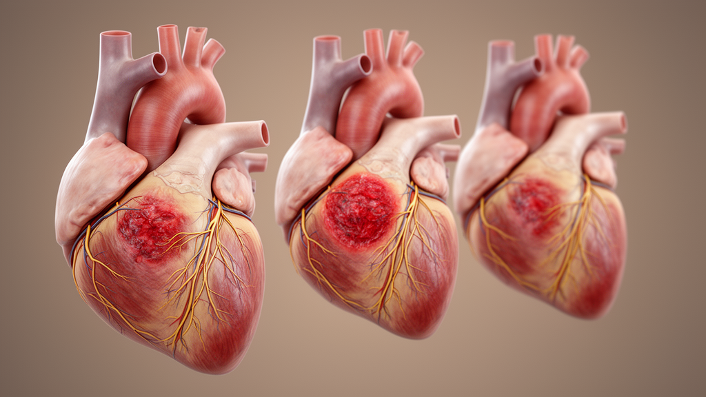
Depletion markers create a parallel interpretive problem. Cardiac troponins T and I are regulatory proteins that leave cardiomyocytes after irreversible membrane damage, with reported loss of immunoreactivity within approximately one to six hours after ischemic onset in working myocardium. Proteomic research has found lower baseline cardiac troponin T and Cx43 expression in nodal tissue than in ventricular myocardium. If normal staining is already faint in a node, it may be difficult to distinguish pathological depletion from constitutive biology or technical variation. The supporting proteomic evidence comes primarily from one published study, however, so the size and reproducibility of these differences remain important subjects for independent research.
Other marker classes may also change their behavior in specialized tissue. Fibronectin, a plasma glycoprotein that accumulates inside cells with damaged membranes, can produce intracellular staining about 30 minutes to four hours after onset in ventricular myocardium. The terminal complement complex C5b-9, also called the membrane attack complex, can label necrotic myocytes as early as one to three hours after ischemia and remain detectable for several days. Neither timing range can simply be transferred to nodes, where abundant collagen, different vascularization and distinct cell biology could alter protein entry or complement activation. Even in ventricular tissue, sensitivity, specificity, observer dependence and post-mortem stability vary among markers, so the published intervals represent estimates of unequal evidentiary strength rather than uniform diagnostic thresholds.
Post-mortem conditions add another unresolved variable. Autolysis may weaken immunoreactivity, and its effect can vary with the duration and conditions of the post-mortem interval. Glycogen-rich Purkinje cells may undergo comparatively early autolytic change, but researchers have not established whether such cellular differences produce meaningful differences in the stability of immunohistochemical markers within the heart. The review does not claim that stability necessarily differs between conduction and working tissue; it stresses that equivalence has never been measured. Forensic interpretation requires the stability of a marker to be known in the tissue actually being examined, making this an evidentiary limitation rather than a demonstrated biological effect.
The stakes are clearest in sudden unexplained death, sudden arrithmic death syndrome and sudden unexpected death in epilepsy. Autopsies in these settings often fail to identify a definite structural or toxicological cause, while the conduction system is considered as a possible site of lethal pathology. A pathologist who finds reduced troponin staining in the sinoatrial node might otherwise infer ischemic injury during the preceding one to six hours using ventricular criteria. But weaker baseline troponin expression could produce a similar appearance without acute injury. Without tissue-specific reference values, the same observation may be physiological, artefactual or pathological, and the difference can affect conclusions about the timing of death, the contribution of a drug overdose or liability for alleged negligence.
Similar caution applies to cases involving diphenhydramine, tricyclic antidepressants, antiarrhythmic drugs and antipsychotic medications associated with conduction-system effects or sudden death, as well as blunt cardiac trauma and procedures such as pacemaker implantation or catheter ablation. Accurate timing could help distinguish acute injury from chronic disease, antemortem trauma from resuscitation artifact or a procedural complication from pre-existing pathology. Yet the review identifies no validated immunohistochemical timing criteria for the conduction system. Current interpretations therefore carry a systematic limitation that should be disclosed whenever such findings contribute to a medico-legal conclusion, rather than being presented as tissue-specific evidence.
The proposed way forward is to treat each part of the conduction system separately. Studies should establish baseline immunoreactivity in non-injured nodes, the bundle of His and Purkinje fibers across age groups and disease backgrounds, then measure changes in controlled ischemic or traumatic lesions with known timing. Post-mortem stability should be tested across the intervals encountered in practice, while investigators examine whether Cx45, Cx40, HCN4 or other proteins preferentially expressed in conduction tissue outperform the Cx43-centered approach used in ventricular myocardium. Standardized dissection and sampling would improve reproducibility, and quantitative image analysis may eventually help assess small, anatomically complex structures. Until such validation exists, the review concludes that ventricular timelines for conduction-system injury are scientifically unvalidated and should be applied with explicit caution.
Subject of Research: Validation of immunohistochemical markers for dating cardiac conduction system injury
Article Title: Immunohistochemical markers for dating cardiac conduction system injury: current knowledge and forensic implications
Article References: Aktar, N., De Simone, S., Fabrello, C., Orsini, F., Bibbò, R., & Cipolloni, L. (2026). Immunohistochemical markers for dating cardiac conduction system injury: current knowledge and forensic implications. International Journal of Legal Medicine. https://doi.org/10.1007/s00414-026-04036-0
Image Credits: AI Generated
DOI: 10.1007/s00414-026-04036-0
Keywords: cardiac conduction system, immunohistochemistry, myocardial injury, forensic pathology, connexin 43, cardiac troponins, sudden cardiac death, injury dating, post-mortem diagnosis, forensic medicine, Immunohistochemical, markers
News Source: Ophelia Keating. (October 22, 2026). Ventricular injury timelines may not fit the heart’s conduction system. Scienmag.