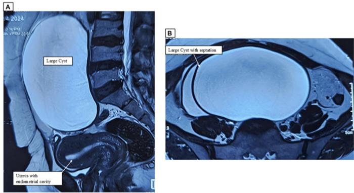

A fascinating case report recently published in Oncoscience showcases the extraordinary co-occurrence of two distinct types of benign ovarian tumors. This unique blend of pathologies, one being a serous cystadenofibroma and the other a collision tumor consisting of serous and mucinous cysts, illustrates the complexities and rarities of ovarian lesions. The case involves a 41-year-old woman who presented with persistent lower abdominal pain and swelling, prompting her medical team to explore the underlying causes through imaging techniques such as MRI.
Upon analyzing the imaging results, the surgical team identified sizeable cystic masses occupying both ovaries. These imaging features raised concerns about potential malignancies, leading to a thorough preoperative assessment. This extensive evaluation included detailed imaging studies and lab analyses to measure tumor markers, ensuring that every avenue was explored to adequately characterize the cystic formations. The discernment shown by the team in evaluating the imaging results underscores the critical nature of preoperative diagnostics, especially in cases where the tumor morphology may closely mimic malignant conditions.
Surgical intervention became necessary after the imaging results confirmed the presence of these unusual masses. In a meticulous operation, the surgical team removed not only the ovaries but also the uterus and fallopian tubes to prevent any complications related to potentially mistaken malignancies. Postoperative pathology results revealed that the tumors were, in fact, completely benign, a conclusion that delighted the patient and her medical team alike. Such outcomes highlight the importance of precise histopathological analysis, which serves as the definitive tool in determining the benign or malignant nature of neoplastic lesions.
Serous cystadenofibromas, the rare terrain navigated by this case, account for only about 1.7% of all benign ovarian tumors. These tumors often go unnoticed during routine gynecological examinations, emerging out of the shadows only when pronounced symptoms arise, such as abdominal distension or discomfort. Moreover, collision tumors, marked by their distinctive coexistence of different tumor types within a singular organ, add another layer of complexity to the diagnostic landscape of ovarian lesions. Their rarity further emphasizes the uniqueness of this case.
For the patient, who had been experiencing symptoms for several months, the journey through diagnosis to surgical intervention proved both challenging and enlightening. Each step taken by her healthcare providers was pivotal, from the initial consultations to the follow-up care that ensured her recovery went smoothly without complications. This successful surgical outcome reinforces the idea that collaboration among specialists, encompassing gynecologists, radiologists, and pathologists, is vital in managing complex cases involving ovarian tumors.
The rarity of such tumor configurations calls for heightened awareness in the medical community regarding their diagnosis and management. Despite their benign classification, the size and appearance of collision tumors can still create an intimidating front, confusing healthcare providers and complicating treatment decisions. The necessity for thorough evaluations cannot be stressed enough, particularly regarding the utility of imaging studies, where misinterpretation could lead to unwarranted surgical procedures.
In the broader field of oncology, this case not only enhances the existing literature regarding serous cystadenofibromas and collision tumors but also serves as a prompt for increased research into uncommon tumor presentations. Each documented case adds to the collective understanding of these complex lesions, inspiring future studies designed to explore their characteristics, treatment modalities, and patient outcomes. As more such occurrences are logged, the medical community moves closer to addressing the enigmatic nature of these tumors effectively.
Sharing unique findings from the surgical realm contributes not only to the academic literature but also to practical applications in clinical settings. The distinctive attributes of this case, including imaging-based features and surgical outcomes, are likely to be discussed at medical conferences, workshops, and other scholarly platforms. Such discourse fosters an environment of learning, where healthcare professionals can refine their skills in identifying and treating unusual tumor presentations.
The case presented here serves as a reminder that every patient represents a unique constellation of symptoms, medical history, and potential tumor profiles. When faced with challenging conditions, the agility of medical professionals in discerning and responding to the nuances of each case can dramatically influence patient care outcomes. In situations laden with uncertainty, such as the presence of unusual ovarian tumors, the meticulous pursuit of knowledge, experience, and collaboration stands as a pillar supporting the foundation of effective healthcare.
Given the unpredictable nature of ovarian tumors, strategies for ongoing surveillance and management post-surgery should be individualized, with considerations for patient history, tumor characteristics, and psychosocial support. As the medical community continues to publish findings and engage in discussions, the hope is that similar cases will circulate, benefiting not only individual patients but also the collective knowledge pool that governs oncological practices.
The remarkable findings from this case serve as a beacon of insight into the intricate world of ovarian neoplasms, emphasizing the necessity for tailored diagnostic and treatment approaches. As research in this domain flourishes, the implications reach far beyond individual case reports, reminding us that every surgical journey holds the potential for enlightening revelation.
This case emphasizes the need for conscientious evaluation and a thorough understanding of pathology, which play critical roles in optimizing patient outcomes, crafting strategic treatment plans, and refining diagnostic techniques in ovarian tumor management.
Subject of Research: People
Article Title: Cystadenofibroma and contralateral collision lesions: A unique ovarian case report
News Publication Date: April 4, 2025
Web References: Oncoscience
References: 10.18632/oncoscience.616
Image Credits: Credit: Copyright: © 2025 Kumar et al.
Keywords: cancer, collision tumor, cystadenofibroma, mucinous, ovary, serous
Tags: benign ovarian tumor co-occurrencecollision tumor in ovariescomplexities of ovarian lesionsimaging techniques for ovarian lesionslower abdominal pain in womenMRI in ovarian diagnosticsovarian tumorspreoperative assessment ovarian tumorsrare ovarian tumor casesserous cystadenofibroma case reportsurgical intervention for ovarian massestumor markers in ovarian pathology


