• HOME
  • NEWS
  • EXPLORE
    • CAREER
      • Companies
      • Jobs
    • EVENTS
    • iGEM
      • News
      • Team
    • PHOTOS
    • VIDEO
    • WIKI
  • BLOG
  • COMMUNITY
    • FACEBOOK
    • INSTAGRAM
    • TWITTER
Wednesday, July 15, 2026
BIOENGINEER.ORG
No Result
View All Result
  • Login
  • HOME
  • NEWS
  • EXPLORE
    • CAREER
      • Companies
      • Jobs
        • Lecturer
        • PhD Studentship
        • Postdoc
        • Research Assistant
    • EVENTS
    • iGEM
      • News
      • Team
    • PHOTOS
    • VIDEO
    • WIKI
  • BLOG
  • COMMUNITY
    • FACEBOOK
    • INSTAGRAM
    • TWITTER
  • HOME
  • NEWS
  • EXPLORE
    • CAREER
      • Companies
      • Jobs
        • Lecturer
        • PhD Studentship
        • Postdoc
        • Research Assistant
    • EVENTS
    • iGEM
      • News
      • Team
    • PHOTOS
    • VIDEO
    • WIKI
  • BLOG
  • COMMUNITY
    • FACEBOOK
    • INSTAGRAM
    • TWITTER
No Result
View All Result
Bioengineer.org
No Result
View All Result
Home NEWS Science News Health

UNC researchers publish striking images of SARS-CoV-2 infected cells

Bioengineer by Bioengineer
September 10, 2020
in Health
Reading Time: 2 mins read
0
IMAGE
Share on FacebookShare on TwitterShare on LinkedinShare on RedditShare on Telegram

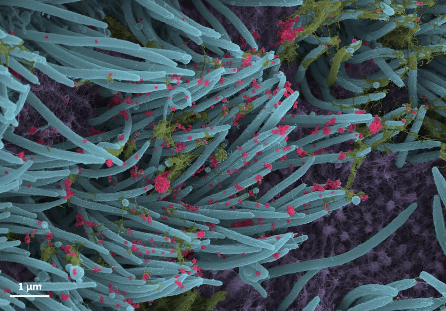
The UNC School of Medicine lab of Camille Ehre, PhD, generated high-powered microscopic images showing startlingly high SARS-CoV-2 viral loads on human respiratory surfaces, ready to spread infection in infected individuals and to others
IMAGE

Credit: Ehre Lab, UNC School of Medicine

The UNC School of Medicine laboratory of Camille Ehre, PhD, Assistant Professor of Pediatrics, produced striking images in respiratory tract cultures of the infectious form of the SARS-CoV-2 virus produced by infected respiratory epithelial cells. The New England Journal of Medicine featured this work in its “Images in Medicine” section.

Ehre, a member of the UNC Marsico Lung Institute and the UNC Children’s Research Institute, captured these images to illustrate how intense the SARS-CoV-2 infection of the airways can be in very graphic and easily understood images. Her lab conducted this research in collaboration with the labs of Ralph Baric, PhD, the William R. Kenan Distinguished Professor of Epidemiology at the UNC Gillings School of Public Health, who holds a joint faculty appointment at the UNC Department of Microbiology and Immunology, and Richard Boucher, MD, the James C. Moeser Eminent Distinguished Professor of Medicine and Director of the Marsico Lung Institute at the UNC School of Medicine.

In a laboratory setting, the researchers inoculated the SARS-Co-V-2 virus into human bronchial epithelial cells, which were then examined 96 hours later using scanning electron microscopy.

The images, re-colorized by UNC medical student Cameron Morrison, show infected ciliated cells with strands of mucus (yellow) attached to cilia tips (blue). Cilia are the hair-like structures on the surface of airway epithelial cells that transport mucus (and trapped viruses) from the lung. A higher power magnification image shows the structure and density of SARS-CoV-2 virions (red) produced by human airway epithelia. Virions are the complete, infectious form of the virus released onto respiratory surfaces by infected host cells.

This imaging research helps illustrate the incredibly high number of virions produced and released per cell inside the human respiratory system. The large viral burden is a source for spread of infection to multiple organs of an infected individual and likely mediates the high frequency of COVID-19 transmission to others. These images make a strong case for the use of masks by infected and uninfected individuals to limit SARS-CoV-2 transmission.

###

The images can be viewed here:

https://www.nejm.org/doi/full/10.1056/NEJMicm2023328?query=TOC

The National Institutes of Health and the Cystic Fibrosis Foundation funded this research.

Media Contact
Mark Derewicz
[email protected]

Related Journal Article

http://dx.doi.org/10.1056/NEJMicm2023328

Tags: Infectious/Emerging DiseasesMedicine/HealthMicrobiology
Share15Tweet9Share3ShareShareShare2

Related Posts

Study finds slight increased risk of sudden vision loss with GLP-1 drugs

July 15, 2026

One-Third of Community Health Centers Still Lack Prenatal Care Services

July 15, 2026

Imaging Study Finds Widespread Brain Connectivity Loss in Schizophrenia

July 15, 2026

Emerging Tick-Borne Virus Sparks Growing Concern

July 15, 2026
Please login to join discussion

POPULAR NEWS

  • New Drug Candidate Developed at McMaster Shows Potential for Treating Brain Cancer

    58 shares
    Share 23 Tweet 15
  • A varied menu

    51 shares
    Share 22 Tweet 12
  • 研究人员开发认知工具包,实现阿尔茨海默症早期检测

    50 shares
    Share 20 Tweet 13
  • Porcine Heart Transplant

    50 shares
    Share 20 Tweet 13

About

We bring you the latest biotechnology news from best research centers and universities around the world. Check our website.

Follow us

Recent News

Study finds slight increased risk of sudden vision loss with GLP-1 drugs

One-Third of Community Health Centers Still Lack Prenatal Care Services

Semipermeable species boundaries allow gene flow but block hybrid swarm in desert fish

Subscribe to Blog via Email

Enter your email address to subscribe to this blog and receive notifications of new posts by email.

Join 85 other subscribers
  • Contact Us

Bioengineer.org © Copyright 2023 All Rights Reserved.

Welcome Back!

Login to your account below

Forgotten Password?

Retrieve your password

Please enter your username or email address to reset your password.

Log In
No Result
View All Result
  • Homepages
    • Home Page 1
    • Home Page 2
  • News
  • National
  • Business
  • Health
  • Lifestyle
  • Science

Bioengineer.org © Copyright 2023 All Rights Reserved.