Necrotizing enterocolitis, or NEC, is one of the most feared emergencies in neonatal medicine. It strikes preterm infants, often within days of starting feeding, and can progress in hours from vague feeding intolerance to transmural bowel necrosis, perforation, sepsis and death. Even with modern intensive care, infants who require surgery face substantial mortality, and survivors carry long-term risks of intestinal failure and neurodevelopmental impairment. Now, a new mini review published in Frontiers in Cell and Developmental Biology argues that two inflammatory forms of regulated cell death, ferroptosis and pyroptosis, may converge to drive the disease, and that this convergence could point the way toward precision-targeted therapies.
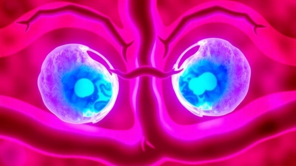
The review, led by Juan He and colleagues at the Affiliated Hospital of Hebei University in Baoding, China, integrates evidence from epithelial, immune and vascular compartments. Ferroptosis is a non-apoptotic, iron-dependent form of cell death driven by catastrophic lipid peroxidation. It occurs when oxidizable phospholipids overwhelm cellular antioxidant defenses, particularly the SLC7A11–glutathione–GPX4 system, which normally detoxifies phospholipid hydroperoxides. ACSL4, an enzyme that enriches membranes with polyunsaturated fatty acids, confers ferroptosis sensitivity. Pyroptosis, by contrast, is a lytic inflammatory death program executed by inflammasome-activated gasdermins. Inflammatory caspases cleave gasdermin D, releasing an N-terminal fragment that forms membrane pores, enabling release of interleukin-1β, interleukin-18 and damage-associated molecular patterns before osmotic rupture.
The premature intestine is developmentally primed for excessive innate immune activation. Epithelial Toll-like receptor 4 signaling impairs enterocyte migration and repair, while reciprocal TLR9 signaling can restrain this response. Breast milk activates epidermal growth factor receptor-dependent protection, and individual human milk oligosaccharides can improve mesenteric perfusion or directly inhibit TLR4 signaling. Maternal aryl hydrocarbon receptor activation also transfers protective signals to the newborn intestine. Importantly, NEC tissue injury is not explained by apoptosis alone; necroptosis has a demonstrable pathogenic role, establishing regulated cell death as a central link between barrier failure and inflammation.
Recent studies provide direct NEC-specific evidence for ferroptosis. Intestinal epithelial farnesoid X receptor promotes ACSL4-dependent ferroptosis; lipid peroxides released from ferroptotic epithelial cells suppress ILC3-derived IL-22, linking epithelial death to defective mucosal repair. FXR antagonism, ACSL4 inhibition and ferroptosis inhibition reduced experimental NEC, while circulating FGF19 and lipid peroxides correlated with disease parameters in infants. Independent transcriptomic work identified ACSL4 as a hub connecting ferroptosis with hypoxia, inflammation and immune-cell abundance. Ferroptosis also occurs outside the epithelium. Inflammatory macrophages from NEC models display iron overload, ACSL4 induction, GPX4 loss and lipid peroxidation; conditioned media from these cells injures epithelial cells, and ferrostatin-1 attenuates both macrophage inflammation and intestinal damage. GPX4-deficient regulatory T cells become ferroptosis-prone in NEC, whereas vitamin E preserves GPX4, Treg viability and immunosuppressive function.
Pyroptotic evidence is equally compelling. Exaggerated TLR4 signaling supplies a strong priming signal for NF-κB-dependent expression of NLRP3 and pro-interleukin-1β, while hypoxia, mitochondrial dysfunction, potassium efflux and microbial products provide activation signals. Cronobacter sakazakii can intensify experimental NEC through TLR4-dependent NLRP3 activation. Pharmacologic blockade of NLRP3 reduces acute intestinal injury and may also limit later neuroinflammatory and cognitive consequences in experimental models, supporting the concept that intestinal pyroptotic signaling can propagate beyond the gut. Sialylated human milk oligosaccharides suppress the TLR4/NF-κB/NLRP3/caspase-1 axis, whereas sodium butyrate reduces NLRP3-dependent GSDMD activation downstream of TLR4/MyD88/NF-κB. Mitochondrial quality control appears to be a critical checkpoint: allicin activates PINK1/Parkin-dependent mitophagy, lowers mitochondrial reactive oxygen species and reduces NLRP3, cleaved caspase-1 and N-terminal GSDMD in epithelial, organoid and neonatal rat models. Pyroptosis may also occur in circulating platelets: GSDMD-dependent platelet pyroptosis promotes exosome release and neutrophil extracellular trap formation through mitochondrial ROS and oxidized mitochondrial DNA, and disulfiram-mediated GSDMD inhibition improves experimental NEC.
Direct reciprocal molecular crosstalk between ferroptosis and pyroptosis has not been demonstrated in human NEC. The evidence instead supports several plausible interfaces through shared stress signals and tissue injury. Ferroptotic lipid peroxides and membrane damage can generate damage-associated molecular patterns, impair epithelial repair and expose the lamina propria to microbial TLR ligands. Mitochondrial ROS and oxidized mitochondrial DNA can simultaneously worsen lipid peroxidation and activate inflammasome signaling. Pyroptotic release of interleukin-1β, interleukin-18 and HMGB1 recruits and activates myeloid cells, increasing oxidative and iron-handling stress. Macrophage ferroptosis and platelet pyroptosis create paracrine signals that injure epithelial and vascular compartments rather than remaining cell-autonomous events. The framework also predicts spatial heterogeneity: ferroptotic epithelium, ferroptosis-resistant surviving cells, pyroptotic immune cells and relatively preserved repair niches may coexist within the same bowel segment, meaning bulk tissue measurements can obscure the cell of origin and the temporal order of events.
HMGB1 is a particularly attractive bridge because it is both a damage-associated molecular pattern released during membrane-disruptive injury and a clinically measurable fecal marker. Fecal HMGB1 rises in early NEC, although current evidence is strongest for suspected or early-stage disease and requires validation across severe surgical phenotypes. Mechanistically, HMGB1 can amplify TLR4/RAGE–NF-κB signaling, thereby linking tissue damage to inflammasome priming; its detectability in stool also makes it more accessible than many research-grade intracellular ferroptosis or pyroptosis markers.
No single biomarker can currently distinguish ferroptosis from pyroptosis at the bedside. Intestinal fatty acid-binding protein reflects enterocyte injury and rises in plasma and urine before or during NEC; higher values can identify complicated disease, but kinetics, renal handling and intestinal maturity affect interpretation. Fecal S100A12 reports phagocyte activation and can rise before severe NEC, although interindividual variability limits stand-alone screening. Reduced Paneth-cell DEFA6, with preserved GUCA2A, suggests impaired antimicrobial defense in resected NEC tissue. Lipopolysaccharide-binding protein has recently shown diagnostic and severity-associated performance, especially when combined with C-reactive protein, but prospective neonatal validation is still needed. A mechanism-informed panel should combine compartments rather than accumulate nonspecific inflammatory tests, including an epithelial-injury marker such as I-FABP, a DAMP such as fecal HMGB1, lipid-peroxidation or ferroptosis-defense measures such as 4-hydroxynonenal, oxidized phospholipids, GPX4 or ACSL4, inflammasome readouts such as cleaved GSDMD, caspase-1, interleukin-18 or ASC specks, and clinical context including gestational age, transfusion exposure, feeding, perfusion and microbiology. Serial sampling is essential because ferroptotic stress may precede overt inflammatory rupture, whereas pyroptotic and NET-associated signals may rise during progression.
Near-term clinical translation should prioritize established supportive measures that reinforce endogenous intestinal protection. Human milk feeding remains the best-supported nutritional strategy; human milk oligosaccharides can promote epithelial regeneration and inhibit TLR4-related inflammatory signaling. Bioactive milk lipids such as dioleoyl phosphatidylethanolamine are mechanistically promising, restoring SLC7A11/GPX4-associated defense and reducing ACSL4-related injury, but dose, formulation and developmental safety remain experimental questions. Ferroptosis-directed approaches include FXR or ACSL4 modulation, restoration of SLC7A11–glutathione–GPX4 activity, iron and lipid-peroxide control, and radical-trapping antioxidants. Ferrostatin-1 remains an experimental NEC intervention supported by preclinical models and is not an approved neonatal therapy. N-acetylcysteine, vitamin E and cystine-related metabolic support have greater clinical familiarity, yet the doses and redox consequences required to alter NEC-specific ferroptosis cannot be inferred from nutritional use. Moreover, indiscriminate antioxidant therapy could interfere with physiological redox signaling and host defense.
Any precision-targeting strategy must preserve the microvasculature and repair niche. Macrophage-derived IGF-1 promotes neonatal intestinal endothelial proliferation and reduces NEC susceptibility, while recombinant IGF-1/IGFBP3 preserves microvascular integrity, enhances epithelial repair and reduces experimental injury. These observations argue for cell-state-specific modulation rather than lineage-wide macrophage depletion: inflammatory macrophage ferroptosis may be pathogenic, whereas trophic macrophage states support vascular growth and epithelial repair. Excessive suppression of inflammasome or myeloid signaling may compromise antimicrobial defense in extremely premature infants. Future research should define the cell-type-specific distribution of ferroptotic and pyroptotic stress across different stages of NEC, particularly within epithelial, myeloid, lymphoid and platelet compartments. Longitudinal, low-volume biomarker panels are needed to determine whether the transition from reversible oxidative stress to self-amplifying inflammatory injury can be identified before irreversible intestinal damage occurs. Spatial transcriptomics, single-cell profiling, lipidomics, neonatal organoid models and multicenter prospective cohorts will be essential for translating regulated-cell-death biology into clinically useful stratification and treatment strategies that interrupt the death–inflammation cycle without impairing host defense or intestinal maturation.
Subject of Research: Cell Death Biology
Article Title: Ferroptosis–pyroptosis crosstalk in neonatal necrotizing enterocolitis: epithelial barrier failure, immune amplification, and precision targeting
Article References: He, J., Gu, Y., An, S., Yan, L., & Song, X. (2026). Ferroptosis–pyroptosis crosstalk in neonatal necrotizing enterocolitis: epithelial barrier failure, immune amplification, and precision targeting. Frontiers in Cell and Developmental Biology, 14, Article 1981457. https://doi.org/10.3389/fcell.2026.1981457
Image Credits: AI Generated
DOI: 10.3389/fcell.2026.1981457
Keywords: necrotizing enterocolitis, ferroptosis, pyroptosis, preterm infants, NLRP3 inflammasome, GPX4, human milk oligosaccharides, biomarkers, crosstalk, neonatal, necrotizing, enterocolitis
News Source: Harold Sullivan. (October 16, 2026). Two Cell Death Pathways May Converge to Drive Deadly Intestinal Disease in Preterm Infants. Scienmag.