


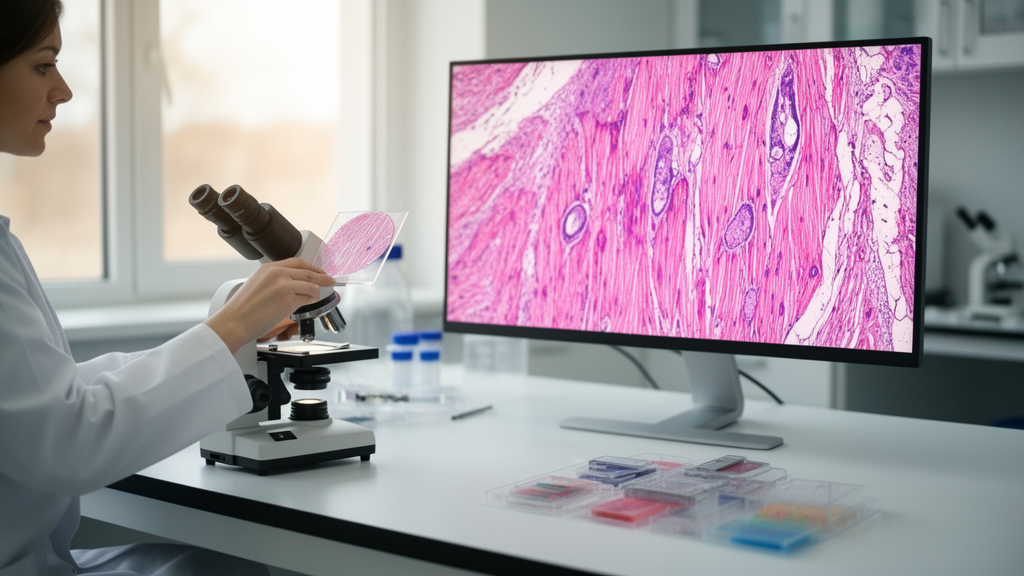
Tissue analysis remains the gold standard for diagnosing a wide spectrum of disorders, yet pathologists perform labor-intensive evaluations to ensure accurate results. A study published in Diagnostic Pathology now describes TopoBoW, a computational framework designed to objectively characterize morphological patterns in microscopy images by capturing both local and global structural features together with their spatial organization. The work addresses a persistent weakness of traditional pixel- or texture-based features, which often fail to represent these different scales of tissue architecture in a single representation.
TopoBoW integrates two complementary image-analysis traditions. Topological Data Analysis, or TDA, captures global structural features of an image, encoding how tissue components are organized across the whole field of view. Bag-of-Visual-Words, or BoVW, encodes local textural information by treating small image patches as discrete visual words whose frequencies describe the microscopic appearance of the sample. By combining these two encodings, the framework builds a feature representation that spans both fine-grained texture and large-scale architecture.
The combined features feed into an attention-guided multi-layer perceptron, a neural network trained to distinguish between healthy and pathological muscle tissue. The attention mechanism allows the model to weight the most informative features during classification. The researchers also used visualizations to examine the statistical behavior of the feature vectors across disease classes and healthy controls, evaluating their discriminative ability rather than relying solely on aggregate performance metrics.
To benchmark the approach, the team compared TopoBoW against several baseline and deep learning models. The comparison set included TDA-based models, histogram of oriented gradients, XGBoost classifiers, attention-based MLP models, and modern convolutional and transformer architectures. Within the image-level cross-validation framework applied to the present preclinical dataset, TopoBoW demonstrated state-of-the-art performance on muscle tissue classification and outperformed all baselines in terms of all classification criteria, including accuracy, F1-score, and AUC.
The study was conducted on mouse models of skeletal muscle disease, a preclinical setting in which histological sections are routinely examined to quantify muscle pathology. Because the framework is interpretable and feature-based, its developers argue that it can assist with pathological research, education, and interactive diagnostic workflows by integrating global structural and local textural information from images. The abstract positions the tool as a support for human experts rather than a replacement for them, consistent with the labor-intensive nature of manual tissue evaluation.
The significance of the method lies in what standard features miss. Pixel- and texture-based descriptors summarize local appearance but can overlook how structures are arranged across an entire image, while purely global summaries can discard fine detail. TopoBoW’s pairing of TDA and BoVW is intended to close that gap, giving classifiers access to both scales simultaneously. The attention-guided MLP then learns which combinations of these features separate healthy from diseased tissue in the training data.
Performance claims in the study are scoped carefully. The reported advantage applies to muscle tissue classification within the image-level cross-validation framework applied to the present preclinical dataset, not to clinical diagnosis in human patients. The article was shared early to provide faster access to peer-reviewed, accepted research; this version is citable and carries a permanent DOI but remains subject to further edits and will be replaced automatically by the final Version of Record.
For researchers working with animal models of muscle disease, the framework offers a way to quantify morphological patterns objectively, reducing the subjectivity and workload of manual assessment. For educators and diagnostic workflow designers, the interpretable feature representation provides a basis for interactive tools in which the features driving a classification can be inspected. The authors describe these applications as the practical payoff of combining topological and textural encodings in a single computational pipeline.
The study, led by Taymaz Akan, Richa Aishwarya, Md. Shenuarin Bhuiyan, and Mohammad Alfrad Nobel Bhuiyan, appears in Diagnostic Pathology as an open-access research article. Its central contribution is methodological: a demonstration that topological signatures and visual word encoding, joined with an attention-guided classifier, can capture morphological patterns in muscle microscopy images more effectively than existing baseline and deep learning alternatives on the tested preclinical data.
Subject of Research: A computational pathology framework that combines topological data analysis and bag-of-visual-words encoding to classify healthy versus diseased skeletal muscle in mouse models.
Article Title: Computational pathology for skeletal muscle disease in mouse models using topological signatures and visual word encoding
Article References: Akan, T., Aishwarya, R., Bhuiyan, M. S., & Bhuiyan, M. A. N. (2026). Computational pathology for skeletal muscle disease in mouse models using topological signatures and visual word encoding. Diagnostic Pathology. https://doi.org/10.1186/s13000-026-01815-6
Image Credits: AI Generated
DOI: 10.1186/s13000-026-01815-6
Keywords: computational pathology, topological data analysis, bag-of-visual-words, skeletal muscle, mouse models, machine learning, image classification, histopathology, attention network, preclinical research, Computational, pathology
News Source: Blake Davidson. (October 30, 2026). Topological Signatures and Visual Words Give Computers a Sharper Eye for Diseased Muscle. Scienmag.



