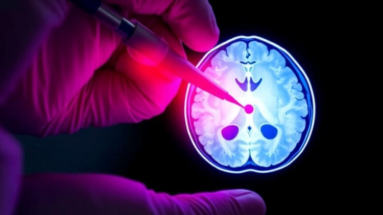
Brain surgeons fighting glioma face one of the most unforgiving challenges in medicine: the tumor does not sit still. Unlike many solid cancers that form a well-defined lump that can be carved away cleanly, gliomas send microscopic tendrils of malignant cells infiltrating into what appears, to the eye and to every instrument in the operating room, to be perfectly healthy brain tissue. Surgeons remove as much of the tumor as they safely can, but the true boundary between diseased and healthy tissue remains invisible during the operation itself. Only after surgery, when pathologists have spent hours analyzing the excised tissue, does anyone learn how close the resection came to capturing the full extent of the disease. A team at Purdue University, working with brain surgeons at the Mayo Clinic, now reports a technique that could compress that waiting period to roughly three minutes, delivering molecular information about the tumor and its margins while the patient is still on the table.
The new study, published in the Proceedings of the National Academy of Sciences, was led by Mahdiyeh Shahi, a former graduate student at Purdue who is now a clinical chemistry postdoctoral fellow at the University of Minnesota Medical School, together with R. Graham Cooks, the Henry Bohn Hass Distinguished Professor of Chemistry in Purdue’s James Tarpo Jr. and Margaret Tarpo Department of Chemistry. The pair collaborated with Dr. Alfredo Quiñones-Hinojosa, a professor of neurosurgery at the Mayo Clinic in Jacksonville, Florida, along with his postdoctoral fellow Loizos Michaelides and other Mayo colleagues, to develop and test a mass spectrometry method designed specifically for the constraints of the operating room. The work draws on an analytical tradition that Cooks himself helped launch more than two decades ago.
Mass spectrometry, at its core, is a way of weighing molecules. The technique ionizes chemical compounds and measures their mass-to-charge ratios, allowing researchers to identify and quantify the complex mixture of metabolites, lipids, and other molecules present in a sample. In a clinical context, that molecular fingerprint can reveal things that conventional microscopy cannot: whether tissue is cancerous, which subtype of tumor it belongs to, and even which genetic mutations the tumor carries. The obstacle has always been speed and practicality. Traditional mass spectrometry requires extensive sample preparation, vacuum systems, and lengthy analysis, none of which fits a surgical workflow in which decisions must be made in minutes.
The Purdue method sidesteps those constraints through ambient ionization, an approach invented at Purdue and first described in 2004. Instead of preparing samples inside sealed vacuum chambers, ambient ionization creates ions from molecules in the open environment, permitting molecular measurements to be made essentially in place. The technique has since been commercialized and widely adopted. The specific variant used in the new study, called syringe touch spray, is strikingly simple: an ordinary hypodermic needle takes a sample from the patient, and the needle itself becomes the ionization source for the mass spectrometric analysis. No elaborate extraction, no separate sampling step, no fundamental redesign of surgical practice.
In the study, the researchers analyzed 86 human brain samples obtained through MRI-guided precision surgery at the Mayo Clinic. During such operations, a small number of samples are taken from surgeon-defined positions in the brain for pathological examination. The team transferred fractions of those samples onto the plunger of a hypodermic needle and sprayed a solution that enabled measurement of the ratio of signals for multiple pairs of endogenous compounds in the brain, using tandem mass spectrometry. The entire measurement took roughly three minutes. Rather than hunting for a single biomarker, the method relies on ratiomics: comparing the relative abundance of pairs of metabolites that naturally occur in brain tissue, which proved far more robust than absolute concentrations that can vary with sample handling.
The results were unambiguous. Ratios of endogenous metabolites distinguished glioma from non-tumor tissue with high confidence, and the same measurements classified tumor subtypes and mutation status, information that normally requires days of laboratory work after surgery. Cooks emphasized that the simple ambient ionization approach enables fast, clinically compatible analysis with the potential to improve margin assessment and enhance patient outcomes. Because glioma cells spread into nearby healthy brain tissue, particularly at the tumor margins, they are notoriously difficult to detect with the conventional technologies available in the operating room. Tissue left behind after surgery can seed recurrence and make subsequent treatment more difficult, so every improvement in margin detection carries direct consequences for survival.
Shahi stressed that the molecular information produced by the technique is simply not available under current standard-of-care procedures. If the diagnostic method were approved for clinical use, she explained, it would provide actionable molecular information during surgery, on a time scale compatible with the operation itself, whereas current pathology results arrive only after hours. That information could directly influence treatment, including the extent of tumor resection, although the final surgical decision would rest with the surgeon weighing the overall clinical context rather than the mass spectrometry results alone. She also noted that the molecular profiles could help guide the selection of targeted therapies, extending the technique’s value beyond the operating room into treatment planning.
Importantly, the researchers are careful to position the technology as an addition to, not a replacement for, existing practice. Cooks noted that the technology would not displace the current standard of care during brain tumor surgery. Instead, it could provide surgeons with molecular information that is not currently available in the operating room, helping to guide decisions during tumor removal, particularly at the tumor margins where the stakes are highest. In previous work published in PNAS in 2024, the team’s measurements were made in the hospital during surgery; in the new study, mass spectrometry data were recorded remotely and compared against the available clinical information, demonstrating that a very simple and quick measurement can carry high discriminating power between diseased and non-diseased tissue while also providing information on tumor grade and mutation status.
The research team included Shahi and Cooks along with Quiñones-Hinojosa; Michaelides; Diogo Moniz Garcia, a former Mayo Clinic postdoctoral fellow now a neurosurgery resident at Washington University in St. Louis; and Aleeshba N. Basil of the Mayo Clinic. Kenneth Lane Virgin of Indiana University School of Medicine, previously affiliated with Purdue’s Aston Laboratories for Mass Spectrometry, also contributed. Shahi and Cooks are members of the Purdue Institute for Cancer Research and work in the Analytical Chemistry group in Purdue’s College of Science. The work was supported by the National Institutes of Health, including the National Cancer Institute’s Innovative Molecular Analysis Technologies Program and the National Center for Advancing Translational Sciences, along with corporate support from Waters Corporation. Specimens came from the Mayo Clinic Florida Neurosurgery BRIDGE Biobank, a central nervous system bioresource built on patient-derived samples.
With the 86-sample study complete, the team’s next step is a similar study involving more than 50 patients in which the technique is used during surgery itself, which Cooks says would pave the way for a clinical trial to be proposed. If those trials succeed, the three-minute molecular readout could become a routine companion to the surgeon’s eye and the pathologist’s microscope, turning one of neurosurgery’s most persistent blind spots into measurable, actionable data at the precise moment it matters most.
Subject of Research: Rapid intraoperative mass spectrometry metabolite ratiomics for glioma margin diagnosis and classification during brain surgery
Article Title: Mass spectrometry offers faster look at the edges of brain tumors
Article References: Mass spectrometry offers faster look at the edges of brain tumors. (n.d.). Original publication
Image Credits: AI Generated
DOI: Not provided
Keywords: glioma, mass spectrometry, brain tumor margins, ambient ionization, syringe touch spray, tumor resection, metabolite ratiomics, intraoperative diagnostics, Purdue University, Mayo Clinic, PNAS, targeted therapy
Cite Scienmag News
APA MLA Chicago
Nathaniel Bowman. (October 1, 2026). Three-minute mass spectrometry test maps brain tumor margins during surgery. Scienmag. https://scienmag.com/three-minute-mass-spectrometry-test-maps-brain-tumor-margins-during-surgery/
Nathaniel Bowman. “Three-minute mass spectrometry test maps brain tumor margins during surgery.” Scienmag, 1 October 2026, https://scienmag.com/three-minute-mass-spectrometry-test-maps-brain-tumor-margins-during-surgery/. Accessed 1 October 2026.
Nathaniel Bowman. “Three-minute mass spectrometry test maps brain tumor margins during surgery.” Scienmag. October 1, 2026. https://scienmag.com/three-minute-mass-spectrometry-test-maps-brain-tumor-margins-during-surgery/
Copy citation Download RIS
Tags: ambient ionizationbrain tumor marginsbrain tumor surgical margin detectiongliomaglioma boundary mapping during surgeryinnovative brain tumor removal techniquesintraoperative cancer tissue characterizationintraoperative diagnosticsintraoperative mass spectrometry for brain tumorsmass spectrometrymass spectrometry in neurosurgeryMayo ClinicMayo Clinic brain tumor surgery advancementsmetabolite ratiomicsPNASPurdue UniversityPurdue University cancer researchrapid molecular diagnostics for brain surgeryreal-time tumor margin analysissyringe touch sprayTargeted therapythree-minute cancer tissue analysistumor infiltration mapping in gliomastumor resection