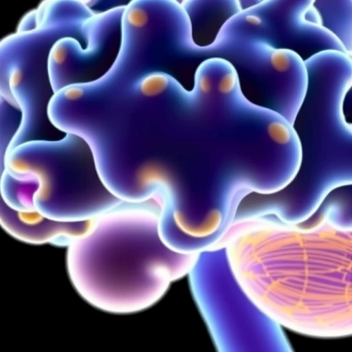
Los Angeles, CA – June 15, 2026 – In a groundbreaking development for brain cancer research, a consortium of leading scientists, spearheaded by researchers at the Terasaki Institute for Biomedical Innovation, has unveiled a comprehensive review that redefines the understanding and classification of glioma organoid models. Published in the prestigious Society for Neuro-Oncology journal, Neuro-Oncology, this exhaustive analysis charts the rapid evolution of glioma organoid technologies and lays out a strategic framework aimed at standardizing research methodologies across the neuro-oncology community.
Gliomas, malignant tumors arising from glial cells in the brain, remain notoriously challenging to study due to their heterogeneity and complex interactions with the brain microenvironment. Traditional two-dimensional models and animal studies have long been limited in their capacity to accurately recapitulate the cellular diversity and three-dimensional architecture intrinsic to human gliomas. This limitation has hindered translational efforts to develop effective therapies. The advent of organoid technology—three-dimensional, human-cell derived tissue cultures—provides an unprecedented opportunity for modeling glioma biology with enhanced fidelity to human disease.
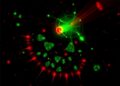
The review article, entitled “Modeling Gliomas with Organoids: Classification, Fidelity, and Guidelines for Translational Neuro-Oncology,” meticulously dissects various approaches to generating glioma organoids. These include engineered organoids built from defined cellular components, patient tissue-derived organoids that retain the native tumor heterogeneity, and assembloids—a novel approach combining multiple organoids to simulate tumor-microenvironment interactions. This layered analysis highlights the strengths and limitations inherent to each model system and underscores the necessity for standardized terminology and classification to bridge gaps in reproducibility and comparability.
Central to the review is the proposal of a three-tier taxonomy for glioma organoid models, designed to refine how researchers categorize and apply these systems. This classification hinges on criteria such as the origin of the cells used, the extent to which the three-dimensional architecture mimics native tissue, and the degree to which critical features like microenvironmental interactions and genetic heterogeneity are recapitulated. By providing a common language and framework, the review encourages a cohesive research ecosystem that facilitates collaboration, data sharing, and comparative analyses.
Beyond classification, the review sets forth evidence-based guidelines to aid investigators in selecting the most appropriate glioma organoid model aligned with specific translational objectives. These guidelines consider factors such as scalability for high-throughput drug screening, fidelity in preserving in vivo tumor characteristics, and capacity for vascularization—critical for mimicking tumor blood supply and therapeutic responses. In doing so, the authors delineate a roadmap that addresses current technological bottlenecks, including challenges in expanding organoids while maintaining cellular complexity and viability.
Research co-leader Zhaohui Wang highlights the timeliness of this work, emphasizing the field’s dynamic yet fragmented nature. “Glioma organoid science is advancing at an astonishing pace, but inconsistent methodologies and terminology have created barriers to collaborative progress,” Wang explains. “Our review aims to unify the community around a structured, shared framework that will catalyze more robust and translationally relevant research.”
The implications of this review extend well beyond academic boundaries. Acting Director of the Terasaki Institute, Dr. Xiling Shen, accentuates the clinical significance, noting that “by harmonizing the standards and accelerating translational applications of glioma organoids, this work serves as a vital link between laboratory innovation and patient-centered therapeutic breakthroughs.”
The intrinsic complexity of gliomas—characterized by cellular heterogeneity, invasive growth patterns, and treatment resistance—necessitates models that truly reflect the intricacies of human pathology. Organoid technology uniquely provides a multidimensional platform where researchers can probe the biology of gliomas in a human-relevant context. This includes the preservation of cell populations such as glioma stem cells, critical players in tumor propagation and resistance, and the replication of tumor niches.
Historically, preclinical models have fallen short by failing to embody tumor microenvironments or genetic diversity, which are pivotal for therapeutic responses. Glioma organoids, with their architecture and cellular heterogeneity preserved, allow researchers to dissect mechanisms of tumor progression and drug resistance with unprecedented accuracy. This positions organoid platforms as transformative tools for predicting patient-specific responses, thus enhancing personalized medicine strategies.
Furthermore, the introduction of assembloids—integrative constructs combining glioma organoids with other brain tissues, such as vasculature or immune components—addresses critical interactions between tumor and microenvironmental elements. This holistic modeling offers opportunities to decode tumor biology within physiologically relevant contexts, enabling investigations into immune evasion, angiogenesis, and metastasis mechanisms at a molecular level.
The review also courageously tackles ongoing challenges in the glioma organoid field, such as scalability hurdles that impede widespread adoption in drug development pipelines. Maintaining organoid viability while achieving vascularization remains another frontier. Strategies involving microfluidics, engineered biomaterials, and co-culture systems are outlined by the authors as promising avenues to elevate organoid complexity and experimental relevance.
Notably, the consortium behind this review includes a diverse array of experts from premier institutions like the University of Pennsylvania, Case Western Reserve University, UCLA, and Emory University, reflecting a truly collaborative spirit aimed at transforming neuro-oncology research paradigms. Emerging scientists included in this collective effort signal a sustained commitment to nurturing innovative research across the glioma organoid landscape.
In sum, this seminal review not only crystallizes the current status of glioma organoid models but also forges a visionary path forward, equipping researchers with the tools and frameworks necessary to harness these technologies for robust translational applications. As gliomas continue to claim countless lives worldwide, this unified approach promises to accelerate the discovery of effective, lifesaving therapies that are urgently needed.
For inquiries and further information, contact Dr. Zhaohui Wang at the Terasaki Institute for Biomedical Innovation via email at [email protected].
Subject of Research: Not applicable
Article Title: Modeling gliomas with organoids: Classification, fidelity, and guidelines for translational neuro-oncology
News Publication Date: June 15, 2026
Web References: http://dx.doi.org/10.1093/neuonc/noag085
Image Credits: Terasaki Institute for Biomedical Innovation
Keywords
Cancer, Neuroscience, Biotechnology, Stem cells, Drug discovery
Tags: 3D glioma tumor modelsbrain cancer research 2026engineered glioma organoidsglioma classification systemglioma heterogeneity modelingglioma organoid models reviewglioma tumor microenvironmenthuman glioma organoidsneuro-oncology organoid technologiespatient-derived glioma organoidsTerasaki Institute glioma researchtranslational neuro-oncology methods

